
Racing the clock
Scientists and patients battle an incurable disease

One morning in May 2006, multiple sclerosis launched its first assault on Antonia Santiago. And a clock began ticking.
Multiple sclerosis is a disabling disease of the central nervous system that affects nearly a million people in the U.S. MS interrupts the flow of information within the brain and between the brain and the body. Symptoms range from numbness and tingling to blindness and paralysis. The progress, severity and symptoms cannot be predicted.
Santiago woke to numbness and tingling in her left leg and foot. A strange burning sensation persisted for days.
“It took a full month to regain all the feeling, to be normal,” she said.
Brain imaging studies and a spinal tap confirmed Santiago’s MS. Although it was a cruel turn for the 20-year-old, she was fortunate to be diagnosed early. Some patients bounce from physician to physician for years with no answer, but Santiago began a weekly injectable therapy right away.
Her journey with MS was just beginning.
***
In 2018 in a laboratory at UT Health San Antonio, a genetic trick gave a young mouse the ability to walk normally. The mouse, bred to lack a gene involved in construction of nerves in the nervous system, had begun to exhibit features of MS disability such as a weakening of the legs and shuffling gait. At 10 months of age, though, the mouse scampered around like a healthy mouse, thanks to a gene rescue strategy.
The gene under study, beta IV-Spectrin, constructs part of the nervous system “highway” through which electrical impulses travel from the brain to muscles. This information highway is under attack in multiple sclerosis. Manzoor Bhat, Ph.D., Zachry Distinguished Chair in Neurosciences, and his team have shown that restoration of the defective gene is more effective if done in mice at 3 months of age rather than later when nerve damage has advanced.
Dr. Bhat believes there is a definite window of time to restore lost nerve function in MS.
***
Santiago’s first seven years with the disease were rough. Flulike side effects from the weekly injection left her in bed 1½ days a week. Thinking she would be better off without therapy, she began to skip doses. In July 2012, an MS flare-up landed her in the hospital.
“I had what seemed like an allergic reaction to an MS medication,” she said. “I broke out in hives at work. It became an exacerbation of the MS, which always starts with numbness.”
She was moved to a rehabilitation hospital that August.
“My walk wasn’t a normal walk; I felt like I was walking on coals,” she said. “And my speech was slowing down.”
Because of the burning feeling, she was afraid to touch anything. Her gait was unsteady, so the therapists gave her a walker and a fall-risk harness. They helped her walk three times a day to bring back sensation in her legs and feet.
“They got me back to walking without assistance,” she said.
Halloween 2012 was her 27th birthday. She spent it in the rehab hospital, and was finally cleared to go home that November.
***
Neurons, the core component of the nervous system, fire electrical impulses that are carried to other neurons and distant muscles. These impulses underlie a person’s cognition, sensory functions and ability to move. The neurons conduct information through axons, which are long, tree trunk-like structures insulated by a protective sheath called myelin. Healthy myelin enables the signals to travel quickly along the axons.
In MS, a person’s own immune system damages the myelin sheath. When the sheath is injured, the signals slow down dramatically, leading to the effects seen in the disease.
But myelin damage is not the end of the story in multiple sclerosis. Dr. Bhat’s team is studying mice that have a deficiency in axonal structures called the nodes of Ranvier. These nodes are gaps in the myelin sheath that function as gatekeepers and amplifiers of the electrical impulses. They, too, enable signals to travel quickly along the axonal highways.
The beta IV-Spectrin gene helps to form and maintain these structures. Mice that lack this gene form immature nodes that fall apart over time. The mice become immobile, and by 6 months of age are essentially paralyzed.
The disease is advancing; the clock is ticking.
***
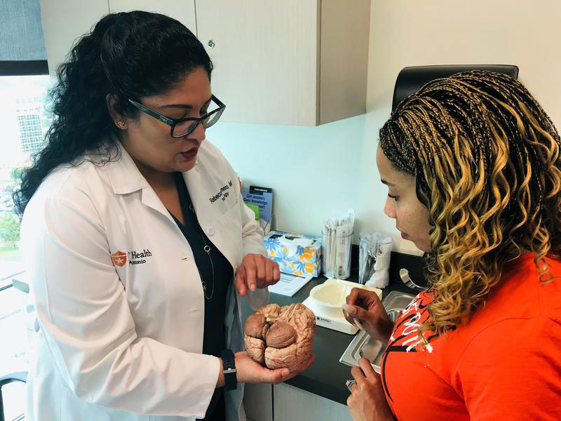
Six years ago, in April 2013, a general neurologist referred Antonia Santiago to Rebecca Romero, M.D., director of the university’s Center for Comprehensive MS Care, a designation from the National Multiple Sclerosis Society. This recognition is presented to centers that offer an MS specialist and integrative, multidisciplinary treatment and care.
Santiago’s long-held wish was to go off injections and start oral therapy. Shortly after her first visit with Dr. Romero, she began taking one of three oral medications approved for MS.
The Center for Comprehensive MS Care offers the latest experimental medications to MS patients in clinical trials.
“The medicines we now have reduce MS progression by 50 percent,” Dr. Romero said. “In 1993 we had our first medication approved in MS, and now we have 14 available medications, and we continue to evolve.”
The future of MS treatment will be in neuro-repair, Dr. Romero said. “That’s the Holy Grail. How do we stimulate our own body to repair the damaged nerves, myelin and axons?”
Breaking down axonal disease into its core components, as Dr. Bhat’s lab is doing, will bring more possibilities into view.
“It’s really important research,” Dr. Romero said. “This is where new therapies will take off.”
***
Since Santiago began seeing Dr. Romero, magnetic resonance imaging (MRI) studies of Santiago’s brain and spine have been stable. She’s had no further hospitalizations for MS flares. Numbness and tingling, however, are constant in the bottom of her feet.
Now 33, she travels around the nation talking to people about MS.
“One of the biggest obstacles to starting treatment is getting diagnosed,” she said. And as Dr. Bhat’s studies show, and outcomes of MS patients themselves reveal, time is critical.
Since the first medication for MS treatment was approved in 1993, “we now have two decades of data on patient outcomes,” Dr. Romero said. “And we know that patients who went on treatment lived longer. We also know that patients who started on medication with the first sign of MS did better long term than those who waited three to six months.”
Identifying a window of time to restore lost function in MS holds promise for future therapies, she said.
“Dr. Bhat recognized that there is a critical time and window in which nerve damage may be repaired. It’s not too far to extrapolate his findings to clinical trials we’ve seen,” Dr. Romero said.
Patients like Santiago live with the knowledge that their MS is progressing. Even in the midst of this, they harbor hope. Because if a mouse’s walking can be restored, there is promise for future MS patients.
“I just know where we were [with MS drugs] when I was diagnosed, and where we are today, and it’s exciting,” Santiago said. “I see a cure coming, maybe in my lifetime.”
To accomplish that, researchers like Dr. Bhat are racing the clock.
‘Zombie’ cells signal death of tissue in Alzheimer’s
A type of cellular stress known to be involved in cancer and aging has now been implicated, for the first time, in Alzheimer’s disease.
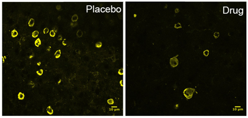
A team of researchers found that the stress, called cellular senescence, is associated with harmful tau protein tangles that are a hallmark of 20 human brain diseases, including Alzheimer’s and traumatic brain injury. The researchers identified senescent cells in postmortem brain tissue from Alzheimer’s patients. Scientists also found them in postmortem tissue from another brain disease, progressive supranuclear palsy.
Cellular senescence allows the stressed cell to survive, but the cell may become like a zombie, functioning abnormally and secreting substances that kill cells around it.
“When cells enter this stage, they change their genetic programming and become pro-inflammatory and toxic,” said study senior author Miranda E. Orr, Ph.D. She is a VA research health scientist at the South Texas Veterans Health Care System, faculty member of the Sam and Ann Barshop Institute for Longevity and Aging Studies, and instructor of pharmacology at UT Health San Antonio. “Their existence means the death of surrounding tissue.” The team reported the discovery in the journal Aging Cell.
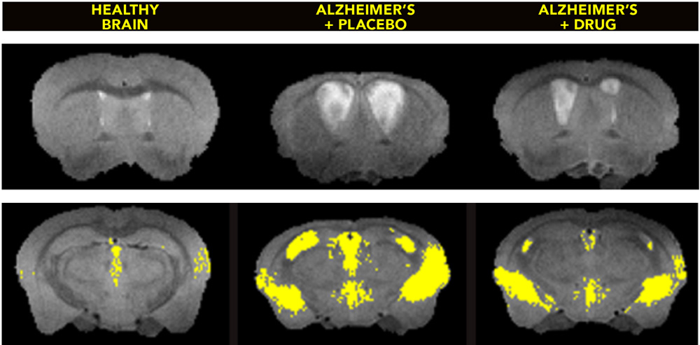
To clear senescent cells from the brains of middle-aged mice with advanced brain disease, researchers used a combination of drugs called senolytics. One drug, dasatinib, is a chemotherapy medication already approved by the Food and Drug Administration. Another drug is quercetin, a flavonoid found in fruits, vegetables and tea.
The combination cleared the senescent cells and led to improvements in brain structure and function—apparently stopping the disease in its tracks.
“The fact we were able to treat very old mice and see improvement gives us hope that this treatment might work in human patients even after they exhibit symptoms of a brain disease,” said Nicolas Musi, M.D., study first author, professor of medicine and director of the Barshop Institute.

Stress might be shrinking your brain
Adults in their 40s and 50s with higher levels of cortisol—a hormone linked to stress—performed worse on memory and other cognitive tasks than peers of the same age with average cortisol levels, research has found.
Higher cortisol in the blood also was associated with smaller brain volumes, according to the study, published in Neurology, the medical journal of the American Academy of Neurology.
“In our quest to understand cognitive aging, one of the factors attracting significant interest and concern is the increasing stress of modern life,” said study senior author Sudha Seshadri, M.D., professor of neurology and founding director of the university’s Glenn Biggs Institute for Alzheimer’s and Neurodegenerative Diseases. “One of the things we know in animals is that stress can lead to cognitive decline. In this study, higher morning cortisol levels in a large sample of people were associated with worse brain structure and cognition.”
The cognitive data are from 2,231 participants in the Framingham Heart Study, for which Dr. Seshadri is a senior investigator; 2,018 participants also underwent MRIs to measure brain volume.
Blood serum cortisol, which varies in level throughout the day, was measured at early morning in each fasting participant. The study featured a relatively young sample of male and female participants.
“Cortisol affects many different functions, so it is important to fully investigate how high levels of the hormone may affect the brain,” said study lead author Justin B. Echouffo-Tcheugui, M.D., Ph.D., of Harvard Medical School. “While other studies have examined cortisol and memory, we believe our large, community-based study is the first to explore, in middle-aged people, fasting blood cortisol levels and brain volume, as well as memory and thinking skills.”
Memory loss and brain shrinkage were found in the study’s middle-age participants before the onset of any symptoms, Dr. Echouffo-Tcheugui noted. He said it is important for physicians to counsel people with higher cortisol levels on ways to reduce stress, such as getting enough sleep and engaging in moderate exercise.
“The faster pace of life today probably means more stress, and when we are stressed, cortisol levels increase because that is our fight-or-flight response,” Dr. Seshadri said. “When we are afraid, when we are threatened in any way, our cortisol levels go up. This study adds to the prevailing wisdom that it’s never too early to be mindful of reducing stress.”
Findings were adjusted for factors including age, sex, smoking and body mass index. The team asked whether having APOE4, a genetic risk factor for cardiovascular disease and Alzheimer’s disease, might be associated with a higher cortisol level. This did not prove to be the case.
The research team included Framingham collaborators at Harvard Medical School; the National Heart, Lung, and Blood Institute; Boston University School of Medicine; the University of California, Davis, at Sacramento; and UT Health San Antonio.

Fighting back against cancer
The Cancer Prevention & Research Institute of Texas awarded more than $2.7 million to fund four projects promoting the fight against cancer at UT Health San Antonio.
Tobacco cessation programs
A $1.3 million grant will enhance tobacco screening and treatment for two groups of patients—those who receive their primary care through the UT Health Physicians medical practice and those who receive their oncology care through
the Mays Cancer Center, home to UT Health San Antonio MD Anderson Cancer Center.
“In addition to providing tobacco cessation services to thousands of patients in these two settings, we will establish a model for innovation in tobacco control delivery that can be readily adopted by other provider systems across Texas,” said Amelie G. Ramirez, Dr.P.H., professor of epidemiology and biostatistics and director of the Institute for Health Promotion Research at UT Health San Antonio.
During patient visits, provider teams will prompt and guide patients who use tobacco to enroll via their smartphones in SMS text messaging or social media direct messaging services.
Preventing HPV-related cancers
In an effort to prevent HPV-related cancers in pediatric cancer survivors, a $1 million, three-year grant was awarded to pediatric oncologist Allison Grimes, M.D., assistant professor of pediatrics and investigator with the Greehey Children’s Cancer Research Institute at UT Health San Antonio.
HPV contributes to more than 30,000 new cancers in the U.S. every year, yet only 43 percent of young females and 32 percent of young males are being vaccinated.
“Childhood cancer survivors represent a particularly high-risk population,” Dr. Grimes said. “Compared to the U.S. general population, pediatric cancer survivors experience significantly higher rates of HPV-related malignancies—40 percent more in females and 150 percent more in males. Despite these increased risks, young survivors have very low HPV vaccination rates. Our project will address this issue across a wide area of the state.”
In South Texas, the majority of children with cancer receive their treatment as part of the South Texas Pediatric Minority Underserved National Cancer Institute Community Oncology Research Program. This network operates as a consortium of five regional pediatric institutions: UT Health San Antonio with clinical partner University Hospital, Methodist Children’s Hospital in San Antonio, Dell Children’s Hospital in Austin, Driscoll Children’s Hospital in Corpus Christi and the Texas Tech University Health Sciences Center in El Paso.
The area for this network encompasses 113 counties and has a population approaching 10 million. The goal is to increase HPV vaccination rates within the network.
Innovative bench research awards
A $200,000 award will support research by Edward P. Hasty, D.V.M., professor of molecular medicine. The BCR-ABL protein, which is generated from the improper fusion of two genes, has unregulated activity that can increase genomic mutations and the risk of cancers including leukemia.
Dr. Hasty will use a new class of drugs developed in his laboratory to fight resistance that arises to drugs that treat cancers expressing BCR-ABL.
CPRIT also awarded $200,000 to a separate UT Health San Antonio research project that targets this protein. Hai Rao, Ph.D., associate professor of molecular medicine, seeks to develop small molecules that would rapidly destroy BCR-ABL.
“The study would lead to new anti-BCR-ABL drugs for patients with chronic myelogenous leukemia and acute lymphocytic leukemia patients,” Dr. Rao said.
Revolutionary new tool could change pancreatic cancer therapies
Pancreatic cancer kills 91 percent of patients within five years of diagnosis. Advances in new therapies have been negligible, and chemotherapies only extend survival by a few months.
A new tool is urgently needed to find a better approach, and Bruno Doiron, Ph.D., assistant professor of medicine, believes he has found one that provides a truer picture of the disease and how it affects humans.
For years, scientists have studied pancreatic cancer by genetically engineering mice to develop the disease or by transplanting tumors into them to test drug activity. The resulting tumors provide an artificial picture of the human disease, Dr. Doiron said.
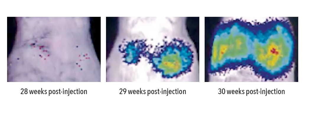
So he and his lab team have found a way to inject a modified virus into healthy adult mouse pancreases. The virus serves as a vehicle for two pro-cancer molecules, present in human pancreatic tumors, to be delivered into the organ. Once injected, the virus permeates the pancreas, yet it doesn’t affect any areas outside of the organ. When the mice reach 28 to 30 weeks of age, tumors develop that resemble human pancreatic cancer.
“This bypasses the artificial manipulation introduced by other methods, and spontaneous cancers develop that mimic those found in people,” Dr. Doiron said.
The lab team uses mice of different breeding and from different parents to ensure the development of the cancer is random, similar to how the disease behaves in humans.
The invention, which has a U.S. patent pending, is significant because “it demonstrates that all previous methods of study are obsolete,” Dr. Doiron said.
This more accurate picture of the human disease could also revolutionize studies of pancreatic cancer initiation and progression, and spur new drug development, said Ruben A. Mesa, M.D., FACP, director of the Mays Cancer Center, the newly named home to UT Health San Antonio MD Anderson Cancer Center.
“This important work by Dr. Doiron and colleagues will allow us to better predict which treatments for the devastating disease of pancreatic cancer will be effective," he said. "These discoveries are a much-needed advance on efforts to cure pancreatic cancer.”

Is stroke in your genes?
A landmark international study of DNA samples from 520,000 people around the world, including 67,000 stroke patients, identified 22 new genetic risk factors for stroke.
It is the largest genetic study of stroke to date, and could lead to stroke drug development, said Sudha Seshadri, M.D., co-author of the study published in Nature Genetics. It also has implications for dementia treatment.
“Understanding these newly identified risk factors for stroke should also enable us to find novel treatments for dementia,” said Dr. Seshadri, founding director of the university's Glenn Biggs Institute for Alzheimer’s & Neurodegenerative Diseases. “Vascular disease in the brain—a series of strokes—can lead to dementia.”
The risk factors identified were for all major subtypes of ischemic stroke, the most common stroke that occurs when a blood vessel supplying the brain is blocked.
The study found the largest correlation between genetic risk factors and blood pressure. Hypertension is a major risk factor for stroke. Vascular health is important for brain function. The brain does not store energy and requires a constant supply of blood and oxygen, as well as blood glucose.
“Any disruption can lead to cognitive problems,” Dr. Seshadri said. “The most obvious example of that is stroke. There is a deficit in the blood supply and that is associated with very obvious changes in cognitive function.”
In another study, Dr. Seshadri and colleagues from the Framingham Heart Study found a trend, over 30 years, of people showing signs of dementia later in life. This is partly attributable to greater control of blood pressure, she said.
“We are looking at other causes, such as lower burden of multiple infections because of vaccination, and possibly lower levels of lead or other pollutants in the atmosphere,” she said. “Early education and nutrition might also play a role.”
Both studies illustrate the strong connection between heart health and brain function.
“What’s good for your heart also seems to be very good for your brain,” she said.

Medical cannabis approved
When a Food and Drug Administration advisory committee in April unanimously approved the first cannabidiol (CBD) medicine for prescription use in epilepsy, it based its decision partly on recommendations of Jose E. Cavazos, M.D., Ph.D., professor of neurology and physiology at UT Health San Antonio.
“I was asked to evaluate the scientific credibility of the evidence—whether there was sufficient research data—to recommend approval of this drug to treat Lennox-Gastaut and Dravet syndromes, which are two types of catastrophic epilepsy,” he said. The syndromes produce multiple seizures daily and treatment options are limited.
CBD is derived from the Cannabis sativa plant, famous as the source of marijuana. CBD is a non-addictive component of the plant, in contrast to tetrahydrocannabinol (THC), which is a component that produces the high of marijuana and is addictive.
The drug, sold under the brand name Epidiolex, is a proprietary oral solution of plant-derived CBD that contains less than 0.1 percent THC and does not produce euphoric effects. Clinical trials indicate the medication can decrease the frequency of “drop attacks” and other symptoms of Lennox-Gastaut. In these attacks, a person suddenly stiffens and falls with the head facing forward. Children with Lennox-Gastaut may wear helmets to prevent head injuries.
Controlled CBD use has been legal in Texas since 2015, when lawmakers passed the Texas Compassionate Use Act. CBD oil dispensaries allow families of children with epilepsy to obtain the oil with physician authorization, but it is a lower dose than the pharmaceutical-grade CBD and is not covered by insurance.
Linda Leary, M.D., clinical associate professor of neurology and pediatrics, said physicians at UT Health San Antonio will assist families who want to try CBD in their children with difficult-to-control epilepsy.
“One reason we are willing to do this is that at least we can help families to use it safely,” Dr. Leary said. “CBD is not viewed as a miracle, but is one more medication that could work to control seizures.”
The FDA decision affects insurance status and provides safety guidelines and dosing instructions.

Chlamydia in your gut could protect you
Exposing the gut to chlamydia protects against subsequent infection in the genital tract and other tissues, researchers have discovered.
Chlamydia is the nation's most common sexually transmitted disease and causes infertility, ectopic pregnancy and pelvic inflammatory disease if untreated.
The protection from exposing the gut to the disease is very robust and is across tissues, which is called transmucosal immunity. Protected sites include the genital tract and the lungs, said Guangming Zhong, M.D., Ph.D., professor of microbiology, immunology and molecular genetics.
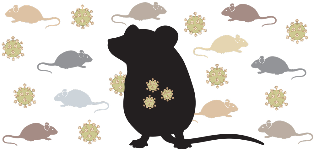
Human exposure to chlamydia is unpredictable, and can come through genital or non-genital sexual contact with an infected partner and perhaps through contact with contaminated materials. The researchers used a mouse model to study the bacteria’s transmission.
They found if the gut was the first site to be colonized by chlamydia bacteria, the mice were immunized against further disease. The gut infection was benign. But if the genital tract was the first to be infected, the resulting disease was harmful, causing a worse disease prognosis, including the possibility of infertility because the disease is advanced before symptoms are present.
The bacterium that causes the disease could be used as an oral vaccine in the future, the researchers believe.
“We take probiotics for our GI health,” Dr. Zhong said. “In the future, we may add chlamydia as a probiotic for the gut. Once the bacteria are established in the GI tract, they don’t spread.”

Stemming the spread of cancer
Scientists at UT Health San Antonio and UTHealth in Houston were awarded $6 million in grants from the U.S. Department of Defense to expand studies of a therapeutic antibody.
The antibody-based drug would be used to stem the spread of breast cancer to bone. This spread, called metastasis, is linked to a dramatic reduction in survival rates.
The lead principal investigator is Jean Jiang, Ph.D., an Ashbel Smith Professor at UT Health San Antonio and the associate director of the Joint Biomedical Engineering Graduate Program of UT Health San Antonio and The University of Texas at San Antonio.
“Antibodies are part of the body’s natural defenses and can be optimized to perform specific tasks,” Dr. Jiang said. “In this case, an antibody activates the connexin channels in bone cells, which protects skeletal tissue from breast cancer colonization and invasion.”
UT Health San Antonio received $3.2 million for preclinical testing in the joint project.
“Research from my laboratory shows the functional role of these channels in suppressing breast cancer invasion and bone metastases. This provides a potential therapeutic target for drug development in breast cancer,” said Dr. Jiang, professor of biochemistry and structural biology.
McGovern Medical School at UTHealth in Houston received $2.8 million for drug development.
The researchers hope to develop a less toxic treatment and reduce deaths tied to the spread of breast cancer to the bone. At the end of the study, they would like to have a drug that can advance to clinical trials.

Research gets a $24 million boost
For the third time in 10 years, UT Health San Antonio has garnered highly competitive National Institutes of Health grants to speed the translation of research discovery into improved patient care.
The university will receive $24 million over the next five years under the Clinical and Translational Science Award Program. The university is a CTSA Program hub and collaborates with eight regional partners, including University Health System, San Antonio Military Health System and The University of Texas at San Antonio.
“This is an enthusiastic vote of confidence in our institution’s ability to affect the future,” said William L. Henrich, M.D., MACP, president of UT Health San Antonio. “We are proud of our track record of accomplishment in advancing community health.”
The program will bring cutting-edge treatments to San Antonio in new clinical trials to affect the most complicated illnesses, said Robert A. Hromas, M.D., FACP, dean of the Joe R. & Teresa Lozano Long School of Medicine and vice president for medical affairs.
“This CTSA Program funding emphasizes maintaining the health of our communities and not just the treatment of illness,” he said.
It will also enable the university to compete for other research awards open only to CTSA institutions, said Andrea Giuffrida, Ph.D., vice president for research and professor of pharmacology.
UT Health San Antonio received its first CTSA designation in 2008 and repeated in 2013.
“We compete with many other prestigious institutions for this funding,” said Robert A. Clark, M.D., MACP, director of the Institute for Integration of Medicine and Science, which administers the CTSA Program at UT Health
San Antonio. “There are only approximately 60 of these CTSA Program hubs throughout the nation, and it’s all the best places.”
Moreover, the university is one of only 43 academic medical centers to function as a CTSA Program hub while at the same time meeting the needs of patients and families through medical practices, including the UT Health San Antonio Physicians practice and the Mays Cancer Center, the newly named home to UT Health San Antonio MD Anderson Cancer Center, a National Cancer Institute-Designated Cancer Center.
CTSA funding does not focus on research of a specific disease but is very broad-based, Dr. Clark said. It supports pilot projects in cancer, heart disease, diabetes, regenerative medicine, aging, neuroscience and a plethora of other areas. Pilot projects are funded based on their potential to mature into major research investigations that can benefit humanity.