
Championing rising investigators
Trustees of the Max and Minnie Tomerlin Voelcker Fund approved more than $2.3 million in new research funding for rising investigators at UT Health San Antonio.
The Voelcker Fund Young Investigator Awards will provide $150,000 a year through 2019 to five faculty. A sixth award of $75,000 will support a one-year pilot study.
The 2017 Voelcker Fund Young Investigator Award recipients are below:
Ann Griffith, Ph.D., assistant professor of microbiology, immunology and molecular genetics, is studying how radiation and chemotherapies damage connective tissue cells in the thymus, a gland that aids in producing immunity. Her project will test whether adding dietary antioxidants during radiation or chemotherapy can boost thymus and T cell recovery.
Zhijie “Jason” Liu, Ph.D., assistant professor of molecular medicine, is studying mechanisms of hormone resistance in breast cancer and the role of estrogen receptor-bound enhancers.
Andrew Pickering, Ph.D., assistant professor of molecular medicine and member of the Sam & Ann Barshop Institute for Longevity & Aging Studies, is studying a gene called TXNRD2 as a novel pharmacological target to improve heart function.
Gangadhara Sareddy, Ph.D., assistant professor of obstetrics and gynecology, studies hormonal and epigenetic signaling involved in the progression of ovarian cancer, breast cancer and glioblastoma. His project will also focus on two epigenetic drugs as a novel therapeutic approach to treat glioblastomas, the deadliest of brain tumors.
Alexei Tumanov, M.D., Ph.D., associate professor of microbiology, immunology and molecular genetics, seeks to understand the fundamental mechanisms of immune regulation of colorectal cancer to develop new immunotherapeutic treatments.
April Risinger, Ph.D., assistant professor of pharmacology, will work on a pilot project focusing on the molecular mechanisms of anticancer drugs called microtubule stabilizing agents.
“The Voelcker Fund medical advisory committee continues to have high praise for the quality of our researchers’ applications,” said Andrea Giuffrida, Ph.D., vice president for research. “This is a credit to our faculty and the scientific excellence of their programs.”
In addition to the Young Investigator Awards, the Voelcker Fund supports the Voelcker Biomedical Research Academy, which provides an immersive biomedical research education and college preparatory program for San Antonio-area high school students, and the Voelcker Biosciences Teacher Academy, which is creating a network of empowered educational professionals who work collaboratively to improve math, science and health education.
Since 2007, the Voelcker Fund has awarded nearly $21 million to fund the university’s research and educational programs.

Meeting a need
The Meadows Foundation has awarded a $100,000 grant to UT Health San Antonio’s Transitional Care Clinic, an innovative psychiatric program operated by the Department of Psychiatry.
The TCC is a short-term clinic that helps people transition from hospital-based psychiatric care to long-term mental health care in the community. The clinic annually treats up to 1,500 patients with serious mental illness until they can find regular care.
The TCC was founded in 2012 as a professional training and clinical care pilot program. It was meant to address the shortage of mental health professionals in San Antonio and the need for quick access to  evaluation, stabilization and short-term care for those transitioning from a hospital to community care.
In its first four years, the TCC provided training in community-based mental health care to 65 medical residents and 150 health professions students. More than 3,000 acutely ill psychiatric patients were served.
It is estimated that in Bexar County alone, 100,000 people suffer from serious mental illness. The shortage of mental health providers and reduced services has resulted in more patients being treated in emergency rooms, which in turn has increased the need for transitional psychiatric care such as the TCC provides.

No more shots
A potential cure for Type 1 diabetes is on the horizon, and the novel approach would also allow Type 2 diabetics to stop insulin shots.
The discovery increases the types of pancreatic cells that secrete insulin.
UT Health San Antonio researchers have a goal to reach human clinical trials in three years, but to do so they must first test the strategy in large-animal studies. Those studies will precede application to the Food and Drug Administration for Investigational New Drug approval, said Bruno Doiron, Ph.D., a co-inventor.
The scientists received a U.S. patent in January, and UT Health San Antonio is spinning out a company to begin commercialization.
The strategy has cured diabetes
in mice.
“It worked perfectly,” said Dr. Doiron, assistant professor of medicine. “We cured mice for one year without any side effects. But it’s a mouse model, so caution is needed. We want to bring this to large animals that are closer to humans in physiology of the endocrine system.”
Ralph DeFronzo, M.D., professor of medicine and chief of the Division of Diabetes, is co-inventor on the patent. “The pancreas has many other cell types besides beta cells, and our approach is to alter these cells so that they start to secrete insulin, but only in response to glucose [sugar],” he said. “This is basically just like beta cells.”
Insulin, which lowers blood sugar, is only made by beta cells. In Type 1 diabetes, beta cells are destroyed by the immune system and the person has no insulin. In Type 2 diabetes, beta cells fail and insulin decreases. At the same time in Type 2, the body doesn’t use insulin efficiently.
The therapy is accomplished by a technique called gene transfer. A virus is used as a vector, or carrier, to introduce selected genes into the pancreas. These genes become incorporated and cause digestive enzymes and other cell types to make insulin.
Gene transfer using a viral vector has been approved nearly 50 times by the FDA to treat various diseases, Dr. DeFronzo said. It is proven in treating rare childhood diseases, and good manufacturing processes ensure safety.
Unlike beta cells, which the body rejects in Type 1 diabetes, the other cell populations of the pancreas co-exist with the body’s immune defenses.
“If a Type 1 diabetic has been living with these cells for 30, 40 or 50 years, and all we’re getting them to do is secrete insulin, we expect there to be no adverse immune response,” Dr. DeFronzo said.
The therapy precisely regulates blood sugar in mice. This could be a major advance over traditional insulin therapy and some diabetes medications that drop blood sugar too low if not closely monitored.
“A major problem we have in the field of Type 1 diabetes is hypoglycemia [low blood sugar],” Dr. Doiron said. “The gene transfer we propose is remarkable because the altered cells match the characteristics of beta cells. Insulin is only released in response to glucose.”
People don’t have symptoms of diabetes until they have lost at least 80 percent of their beta cells, Dr. Doiron said.
“We don’t need to replicate all of the insulin-making function of beta cells,” he said. “Only 20 percent restoration of this capacity is sufficient for a cure of Type 1.”
Staying in rhythm
There’s a new technique UT Health San Antonio physicians are using to treat atrial fibrillation, a common heart rhythm disorder and major risk factor for stroke.
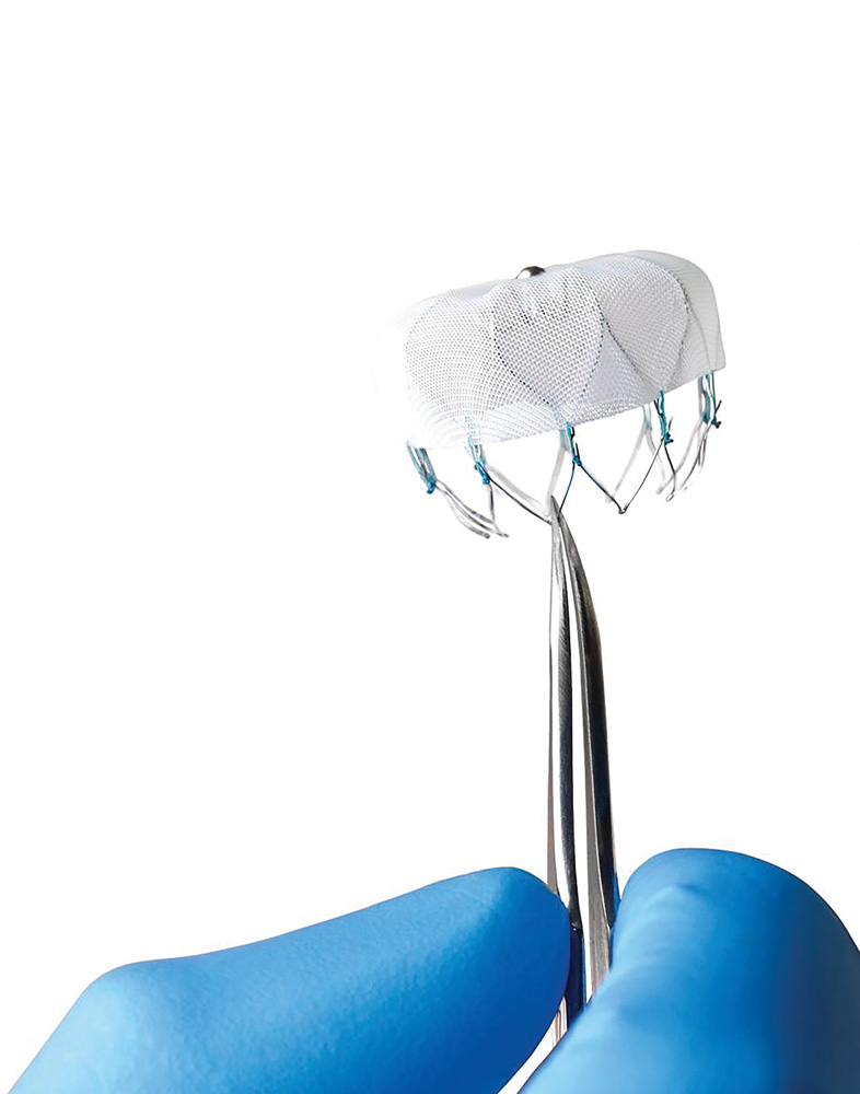
They are among the first in Texas trained to implant the Watchman™ left atrial appendage closure device, which is approved by the Food and Drug Administration as a therapy for symptomatic, persistent atrial fibrillation.
Atrial fibrillation, known as afib, affects around 6 million people in the United States. It is an irregular heartbeat that can result in very life-limiting symptoms for many patients. Although it occurs in both young and old people, the prevalence of afib increases with age. A serious consequence can be a stroke, which is up to five times more likely in patients with afib.
“Atrial fibrillation continues to be a very challenging problem to treat,” said Steven R. Bailey, M.D., professor and chief of the Janey and Dolph Briscoe Division of Cardiology. “The prevalence of afib is growing, and there is no cure for the disease. However, it is hoped that studying new strategies may result in more effective ways of treating our patients who are struggling with afib. It is our goal to improve the quality of life of our patients and to limit the adverse events related to atrial fibrillation.”
The Watchman procedure closes a pouch called the left atrial appendage.
“Although the left atrial appendage is a small structure, it is believed to play an important role in the initiation of afib,” said Manoj Panday, M.D., director of cardiac electrophysiology and an associate professor of medicine.
The left atrial appendage is the source of up to 90 percent of strokes that are caused by afib.
“Patients have to be on lifelong blood thinner to reduce the risk of clots forming in that small area of the heart,” Dr. Panday said.
The Watchman device is delivered via a special catheter. The device is a self-expanding, nickel-titanium frame with an attached woven plastic cap that, when open, looks like a jellyfish. The device is positioned and then expanded to close blood flow to the left atrial appendage. Within six weeks endothelial tissue grows over the device, which remains for life.
“Once implanted successfully, the device eliminates the need for long-term oral anticoagulation, which is especially important for those who are not eligible to have blood thinners,” Dr. Panday said.

Zero pain
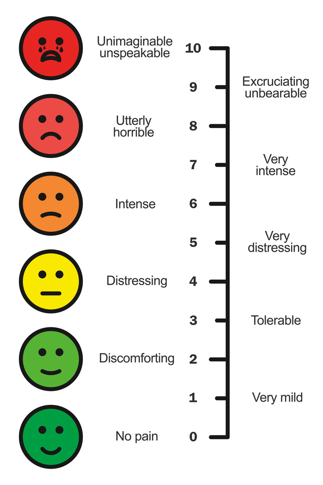 What if there were a safe and effective way to control pain at its source, tailored to each patient, yet without the risk of addiction and overdose?
Mark S. Shapiro, Ph.D., professor of physiology, said unexpected and provocative results from his research indicates this could someday become a reality.
The key lies in our ions. Charged salt atoms, or ions, such as calcium and sodium, pass in and out of cells through protein pores called ion channels. Dr. Shapiro and a team of researchers discovered that two ion channels in sensory nerve cells are interacting in unexpected ways. Sensory nerve cells are neurons that initiate the sensations of pain or burning.
“One ion channel we studied, called TRPV1, starts the signal that tells the brain something hurts,” he said. “The other channel, a calcium ion channel called CaV1.2, signals genes to turn on or off in response to activity, such as painful stimuli.”
The researchers saw that proteins of these ion channels interact with each other and function in tandem.
“It was thought that those two processes occurred in different parts of the neuron,” he said.
Ion channels are in the outer membranes of all cells, including neurons, the cells of the nervous system. The finding that these two channels are coupled has important implications for all neurons, including sympathetic neurons, which control automatic functions of the body such as heart rate and breathing, and sensory neurons, which sense painful stimuli and sound the alarm to the brain.
“The most unexpected, exciting and provocative result of this work is the discovery that the ion channel that starts the pain signal is intimately associated with the ion channel that turns on or off genes’ response. They are locked together,” he said. “It’s as if two people are in locked arms, and when one takes a step, the other moves in lockstep.”
The researchers will now study whether turning genes on or off can stop the pain cycle that is often so debilitating. It theoretically should be possible to reduce the association between the ion channels to lower the pain signal and remove such chronic pain. In individuals who have lost pain sensation in part of the body, such as in the feet, it might be possible to increase the signal. This would increase sensitivity to pain to help them avoid injuries such as burns or ulcers.
“All we have now are centrally acting opioid painkillers—fentanyl, hydrocodone and others—which has led to an epidemic of abuse and overdoses,” Dr. Shapiro said. “These medications don’t stop the pain signal but instead cover up the sensation in the brain, which frequently leads to devastating addiction. We want to treat pain at the source, at the sensory neuron, so that the pain signal never gets started in the first place, or if it does get started, doesn’t lead to this vicious cycle of pain and addiction.”
The results of his study were reported in the journal Neuron.

Going molecular
We know that exercise benefits the mind and body, but we don’t know how it affects us at the molecular level.
So UT Health San Antonio and UT Medical Branch at Galveston are teaming up to study how exercise changes the body at the smallest level. The project is funded through $4.5 million from the National Institutes of Health over the next six years. The funds are the first from the NIH Common Fund for its Molecular Transducers of Physical Activity in Humans Consortium. They were distributed to the Texas team and other groups nationwide.
“We have long understood that exercising is beneficial to our overall health, but we don’t fully understand the impact of exercise at the molecular level,” said NIH Director Francis S. Collins, M.D., Ph.D. “The development of a so-called molecular map of circulating signals produced by physical activity will allow us to discover, at a fundamental level, how physical activity affects our health. This knowledge should allow researchers and doctors to develop individually targeted exercise recommendations and better help those who are unable to exercise.”
A team led by UT Health San Antonio’s Nicolas Musi, M.D., and Blake Rasmussen, Ph.D., at the UT Medical Branch at Galveston, is one of six groups that will recruit healthy adults for an exercise study. The investigators will collect blood, urine and tissue samples from active and sedentary volunteers who will perform resistance or aerobic exercises. These samples will be shared with colleagues at the consortium’s Chemical Analysis Sites.
“Previous research by my laboratory has centered on the effects of exercise on the mitochondria, which function as the power plants of the cells,” said Dr. Musi, professor in the Joe R. & Teresa Lozano Long School of Medicine and director of the Barshop Institute for Longevity and Aging Studies.
“Exercise increases the size and number of the mitochondria. Information gleaned from this new study funded by NIH will further enhance our understanding of the physiological benefits
of exercise.”

Stop the music
 Our genes are the blueprints of our lives. They determine how tall we are, whether we have freckles and the color of our hair. They also play a role in our health, influencing whether we are well or sick. Some diseases can be traced to defects in genes, but, enigmatically, not all can. Brothers and sisters with very similar genetic makeups may have vastly different health outcomes at life’s end. Why?
An emerging area of science—epigenetics—offers an answer. In fact, a School of Medicine researcher’s studies may demonstrate that an epigenetic link exists between two of the most common diseases of our time.
Epigenetics is the study of biological processes that switch genes on and off without altering the genetic code itself. Epigenetic changes may be prompted by the environment, diet, stress, aging and other factors.
Of particular interest to Kexin Xu, Ph.D., assistant professor of molecular medicine at UT Health San Antonio, is that epigenetic changes are reversible. That means that some of the world’s most prolific and damaging diseases, such as cancer and diabetes, may not just be halted. The diseases themselves may be reversed.
“By stopping undesirable biological processes that promote cancer, diabetes or both, we can alter the lives of hundreds of thousands of people suffering the effects of these diseases,”
Dr. Xu said. “Epigenetic reprogramming has the potential to do that.”
It’s a common analogy in the science world: Epigenetics is like an invisible piano player hovering over our genetic keyboard, with each key representing a different gene. The pianist controls which keys get pressed, the rhythm and the melody. But what if the musician can be trained to play a different melody? Trained to stop striking the keys that represent diseases such as cancer or diabetes?
As scientists such as Dr. Xu learn more, it’s conceivable therapies will be developed to “stop the music” and replace an off-key melody with a beautiful harmony.
“There is a close association between diabetes and cancer,” Dr. Xu said. “We are focused on a pair of epigenetic programs that could be changed, resulting in treatment of one or both diseases.”
The body’s energy usage is called metabolism. Studies have shown that impaired metabolism, a hallmark of diabetes and obesity, plays an important role in cancer development and progression. For example, the National Institutes of Health estimates that 20 percent of cancer deaths are associated with obesity.
To examine this linkage, Dr. Xu has her eye on a destructive alliance between a pro-cancer protein called EZH2 and a metabolic pathway that acts as a sensor of cell energy, O-GlcNAcylation. A defect there may be the culprit.
The pro-cancer protein EZH2 drives the aggressiveness of cancer cells and may interfere with the energy sensor O-GlcNAc’s function as a metabolic thermostat.
“We want to target the cross-talk of these two and try to cure both cancer and diabetes by making epigenetic changes,” Dr. Xu said.
By regulating the two, it may be possible to reverse the situation to the normal state.
“Kexin’s work has the potential to improve cancer treatment,” said Tim Huang, Ph.D., professor and chairman of molecular medicine. “Epigenetic reprogramming could eliminate drug resistance in cancer patients who are not responding to therapies.”
Once again using the piano player analogy, by prompting the musician to play a different tune or style, a revolutionary cancer drug—or revolutionary diabetes drug—could result.
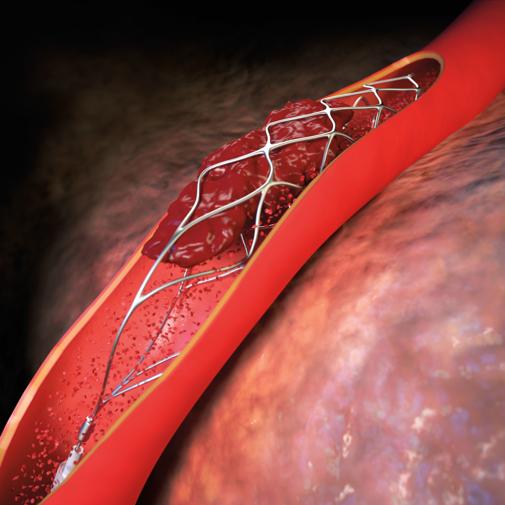
Stents for strokes
After 61-year-old Bobbie Thompson had an ischemic stroke, her face drooped. She couldn’t lift her left arm. Her carotid artery carrying oxygen-rich blood to her brain was blocked.
Without immediate intervention, her brain tissue would begin to die.
Each year, nearly 800,000 people in the U.S. have a stroke. About 87 percent of the time, the cause is a clot that blocks an artery and restricts blood flow to the brain, called an ischemic stroke. Because brain tissue begins to die, opening the artery quickly is essential.
For 20 years, physicians have used the clot-busting medication called intravenous tPA, but often it doesn’t help patients, like Thompson, whose largest vessels are blocked. They are now pairing emergency interventional procedures to open the vessel, save brain tissue and preserve quality of life.
Using devices called “stent retrievers,” they extract clots from stroke patients with large-vessel blockages. Stent retrievers, about the size of a coffee stir, are attached to a catheter that is threaded from the groin to the brain’s largest vessels. The stent is used to open a hole through the clot and reestablish blood flow. The stent attaches to the blood clot and is then pulled back down the vessel toward the groin and removed from the body.
Restoring blood flow as quickly as possible is important in most stroke patients with large-vessel blockages who are transported to the hospital early, said neurosurgeon Ramesh Grandhi, M.D., assistant professor in the Department of Neurosurgery.
“Amongst those patients, CT imaging shows that a large portion of the brain is at risk of dying from a blood vessel blockage but is not yet dead,” Dr. Grandhi said. “If circulation resumes quickly enough, the at-risk brain survives.”
In cases such as Thompson’s, intravenous tPA may not work by itself. The resulting stroke can cause patients to lose so much function that they require around-the-clock nursing home care.
But with the help of stent retrievers, a full recovery is possible. Two days after her stroke, Thompson had no residual paralysis. Today, she smiles fully, can move both arms freely, and walks around without a limp.
“Her arm would never have moved again, but here she is giving a thumbs-up to her doctors,” said neurosurgeon Jeremiah Johnson, M.D., of UT Medicine. “Those are the kind of results we want for our stroke patients.”
The UT Medicine Comprehensive Stroke and Cerebrovascular Program includes four vascular neurologists, three neuro-interventional surgeons, a neuro-intensive care team, rehabilitation medicine specialists and additional neurosurgeons. The team operates at University Hospital and in the Baptist Health System.
“UT Medicine provides the community a comprehensive team of academic experts who are supported by our clinical partners,” said David F. Jimenez, M.D., FACS, professor and chairman of the Department of Neurosurgery. “From stroke to brain aneurysms to other conditions, there isn’t anything in the cerebrovascular world that we don’t do here.”

Walking time bomb
It’s an unsettling thought: You could be walking around for 20 years developing Parkinson’s disease and not even know it.
And once symptoms appear, it’s too late for a cure.
What if a therapy that treats the root causes of Parkinson’s, not just the symptoms, could be started earlier?
Researchers in the School of Medicine are studying changes in Parkinson’s-affected cells at various stages of the disease, long before any symptoms are evident. They describe the changes in the Journal of Neuroscience.
The hope of the research is twofold: gain understandings that can be used to formulate a drug to arrest the disease at a halfway point, and lengthen the time when patients with Parkinson’s can lead healthy, productive lives.
“For the first time, we are getting a look at what’s going on in the time window before the disease visibly takes hold but while changes are occurring,” said study senior author Michael Beckstead, Ph.D., assistant professor of physiology and a member of the Barshop Institute for Aging and Longevity Studies.
Parkinson’s is marked by the degeneration and death of cells called dopamine neurons. These neurons are found in a brain structure called the substantia nigra. The researchers studied mice in which only these neurons are affected by a genetic mutation.
In the mice, mitochondrial activity is hampered just in dopamine neurons of the substantia nigra. Mitochondria produce energy for our cells, and since these mice have impaired mitochondria, their dopamine neurons don’t make energy efficiently.
At first, the mice are completely normal, but as weeks and months go by, the mutation causes their dopamine neurons to slowly become sick and die off.
“It’s a progressive model in that these changes don’t take place overnight,” Dr. Beckstead said. “This makes it like the human disease, which is thought to be somewhere in the range of a 20-year process before symptoms become evident.”
In the mice, behavioral symptoms such as tremors start to manifest when the mice are about 20 weeks old. The study assessed functional status at time points before that—comparing dopamine neuron function at 6 to 10 weeks of age with function at 11 to 15 weeks of age and function at 16-plus weeks.
With these comparisons, the researchers constructed a timeline of functional decline in the dopamine neurons. They observed smaller dopamine neurons, reduced communication between the neurons and impaired electrical activity of the neurons.
“Pretty much everything we measured declined in these cells,” Dr. Beckstead said. “It was really remarkable how everything we studied changed. It was a general decline, and these changes were all occurring before the animals were symptomatic, before you could detect any sort of deficit in their movement.”
In older mice starting to display the abnormal movements of the disease, the scientists made another observation—heightened gene expression to increase the electrical activity in the dopamine neurons.
“This is a late occurrence in the disease process,” Dr. Beckstead said. “We believe the cells are trying to compensate for the declining electrical activity. That’s probably how humans are able to be free of symptoms for so long when they have Parkinson’s, even though 30 percent or more of their dopamine neurons have died out.”
The study results aren’t going to translate into a clinical therapy any time soon, but such findings offer the promise that one day the root cause of Parkinson’s may be understood and treated.
Current treatments for Parkinson’s disease are all symptomatic. They focus on improving the movement deficits and making the patients more comfortable.
“We don’t have any treatments right now that actually affect the disease process,” Dr. Beckstead said. “The reason we don’t have any is we don’t understand what’s going on in the early stages of this disease. Studies such as ours will help fill in those knowledge gaps.”
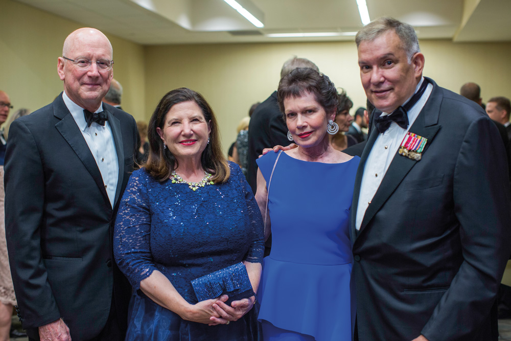
A salute to service
UT Health San Antonio’s historic ties with the military and the life-changing collaborations between the two were celebrated at the ninth annual President’s Gala in September.
More than 1,600 community leaders, faculty, staff and students attended the gala that raised over $500,000. William L. Henrich, M.D., MACP, university president, announced that gala proceeds will establish The Maj. Gen. (USA Ret.) Joe and Patty Robles Chair in Military Health Research.
Gen. and Mrs. Robles were recognized for their service to the nation, San Antonio’s military community and USAA. Gen. Robles served USAA as president and CEO from 2007 until his retirement in 2015. UT System Chancellor Bill McRaven, a retired U.S. Navy four-star admiral, and his wife, Georgeann, served as honorary chairs of the gala.
“Joe and Patty have made, and continue to make, exemplary contributions to San Antonio, to our military and to the welfare of our entire community and country,” Dr. Henrich said. “They are our city’s finest examples of integrity and kindness, and they embody military values in all respects.”
The Robles Chair in Military Health Research, Dr. Henrich added, “will be dedicated in perpetuity to supporting [UT Health San Antonio] as we lead innovative medical research, health education and clinical care that enhances and nurtures military collaborations. One of our top priorities is to improve the health and well-being of military personnel and veterans throughout our community, state and nation.”
Gen. Robles was drafted into the U.S. Army in 1966 and served in multiple command and staff positions, including active-duty posts in Korea, Vietnam and Germany, and in Operations Desert Shield and Desert Storm. He retired in 1994 and joined USAA that year, serving as chief financial officer and controller and in other executive administrative positions.
The university has enjoyed a close relationship with the military for decades. In 2014, UT Health San Antonio formalized this collaboration by establishing the Military Health Institute under the leadership of Byron C. Hepburn, M.D., Maj. Gen. (USAF Ret.), founding director of the institute and associate vice president of UT Health San Antonio.
According to its vision statement, the Military Health Institute will be the nation’s leader in health-related academic collaborations that improve the health and resiliency of the nation’s military service members, veterans and their families.