A type of cellular stress known to be involved in cancer and aging has now been implicated, for the first time, in Alzheimer’s disease.
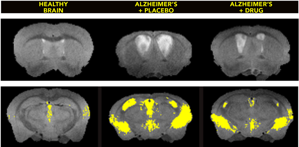
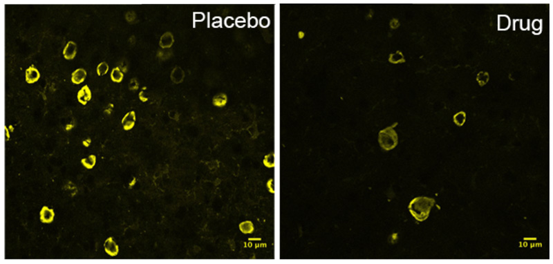
A team of researchers found that the stress, called cellular senescence, is associated with harmful tau protein tangles that are a hallmark of 20 human brain diseases, including Alzheimer’s and traumatic brain injury. The researchers identified senescent cells in postmortem brain tissue from Alzheimer’s patients. Scientists also found them in postmortem tissue from another brain disease, progressive supranuclear palsy.
Cellular senescence allows the stressed cell to survive, but the cell may become like a zombie, functioning abnormally and secreting substances that kill cells around it.
“When cells enter this stage, they change their genetic programming and become pro-inflammatory and toxic,” said study senior author Miranda E. Orr, Ph.D. She is a VA research health scientist at the South Texas Veterans Health Care System, faculty member of the Sam and Ann Barshop Institute for Longevity and Aging Studies, and instructor of pharmacology at UT Health San Antonio. “Their existence means the death of surrounding tissue.” The team reported the discovery in the journal Aging Cell.
To clear senescent cells from the brains of middle-aged mice with advanced brain disease, researchers used a combination of drugs called senolytics. One drug, dasatinib, is a chemotherapy medication already approved by the Food and Drug Administration. Another drug is quercetin, a flavonoid found in fruits, vegetables and tea.
The combination cleared the senescent cells and led to improvements in brain structure and function—apparently stopping the disease in its tracks.
“The fact we were able to treat very old mice and see improvement gives us hope that this treatment might work in human patients even after they exhibit symptoms of a brain disease,” said Nicolas Musi, M.D., study first author, professor of medicine and director of the Barshop Institute.