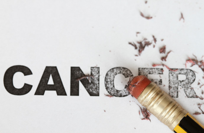
Women scientists lead efforts to fight cancer
Vivienne Rebel, M.D., Ph.D., assistant professor of cellular and structural biology, and Gail Tomlinson, M.D., Ph.D., professor of pediatrics, are two women in science who play crucial roles at the Greehey Children’s Cancer Research Institute. Dr. Rebel dedicates her time to research that could one day translate to new therapies, while Dr. Tomlinson cares for seriously ill children and seeks to translate research discoveries.
Dr. Tomlinson, who holds the Greehey Distinguished Chair in the Genetics of Cancer, is division chief of hematology-oncology. She also holds the Greehey Distinguished Chair for the Children’s Cancer Research Institute Director at the Greehey Institute where she serves as interim director. She sees hospitalized children and has a grant from the Cancer Prevention and Research Institute of Texas (CPRIT) to study pediatric liver cancer. This work is developing into a national and international clinical research trial.
Dr. Rebel investigates the properties of stem cells in a bone marrow disease called myelodysplastic syndrome (MDS). "Stem cells are the only cells in the body that have a seemingly unlimited capacity to proliferate, just like cancer cells," she said. "I thought that by studying stem cells, we may learn about cancer." Dr. Rebel’s laboratory is using a mouse model to try to understand what is going wrong in the production of blood-forming stem cells that may eventually lead to MDS and, in some cases, to leukemia. "It is thought that the culprit cell of MDS is the blood-forming stem cell," she said.
Dr. Tomlinson became intrigued by the molecular basis of cancer while a biochemistry student at Duke University. "I was interested in how some of the biochemical findings could influence the cure of pediatric diseases such as leukemia," she said. "After I became more established, I focused my efforts on understanding genetic causes of pediatric tumors, all with a translational goal in mind to help guide therapies or understand causes so that, whenever possible, diseases could be prevented or detected early. Most pediatric cancers have historically not been thought to be preventable. It is a goal, albeit a long way out."

Dr. Tomlinson is principal investigator on a $2.7 million CPRIT grant to empower health-care providers to map out cancer risks of their patients and to share information about family history as an important factor. The grant, awarded in 2012, also supports screening services for people at high risk for cancer who may not have adequate access to screening.
Dr. Rebel admires women scientists such as Bettie Sue Masters, Ph.D., the Robert A. Welch Foundation Distinguished Chair in Chemistry at the Health Science Center. "Dr. Masters is amazing," Dr. Rebel said. "She has faced the real difficulty of being from a generation when being a woman scientist wasn’t easy. Over time, there has been improvement." Dr. Rebel also greatly respects Dr. Tomlinson for juggling both a clinic schedule and the institute. "I decided to stay with research and not go into the clinic," she said.
As these two women apply their intellect, compassion and resources to understanding pediatric cancers, the children of Texas and the world are the beneficiaries.

Using an infant virus to fight cancer
School of Medicine researchers’ discovery proves effective as cancer treatment
Discoveries in science sometimes come by serendipity, when hard and consistent work done in one area of interest leads to a stunning discovery in a related or even unrelated area. Santanu Bose, Ph.D., associate professor of microbiology and immunology in the Joe R. and Teresa Lozano Long School of Medicine at the Health Science Center, knows by experience that this can happen.
Dr. Bose was studying the immune response of normal and cancerous cells to RSV (respiratory syncytial virus), which causes respiratory infections in infants and young children. Without specifically looking for a cancer treatment, he saw that the virus was "oncolytic" - it preferentially infected and damaged cancer cells while leaving the healthy cells alone. This propelled a new line of research. Dr. Bose teamed with Bandana Chatterjee, Ph.D., of the Long School of Medicine and the South Texas Veterans Health Care System, to test RSV in her mouse model of prostate cancer. Those results again showed a robust anti-cancer effect of RSV. Mice with prostate tumors were treated with the virus and within a week the tumors were gone. "We kept the mice for four months, and the tumors never came back," Dr. Bose said.
Now Dr. Bose is the inventor on a pending U.S. patent of RSV as an oncolytic therapy. This represents a new use for the virus. Dr. Chatterjee, professor of molecular medicine, is the co-inventor. CZ BioMed Corp. of Tampa, Fla., licensed the oncolytic use of RSV in an agreement with South Texas Technology Management (STTM), a regional University of Texas technology-transfer office managed by the Health Science Center. RSV is already showing effectiveness in human trials abroad, according to a company statement.
Dr. Bose, whose work is funded by the National Institutes of Health, said, "This is an exciting development because this is a homegrown invention that is being tested in humans, and therefore this scientific discovery has direct clinical, translational relevance."
"We are pleased that CZ BioMed has agreed to work with us to commercialize Dr. Bose’s and Dr. Chatterjee’s exciting discovery to efficiently target and treat different forms of cancer," said STTM Executive Director Arjun Sanga, J.D., assistant vice president for technology transfer at the Health Science Center.
Dr. Chatterjee said it is significant that the virus killed tumors even in mice with competent immune systems. This mirrors human patients who have functioning immune defenses. RSV also worked whether it was injected directly into the tumor or systemically through the abdomen. "This is important because there are some tumors to which you can inject the drug directly, whereas others you can’t and a drug must work systemically," Dr. Chatterjee said.
Her work on the RSV project is funded by a Merit-Review grant from the U.S. Department of Veterans Affairs (VA), a VA Senior Research Career Scientist Award, and a grant to Drs. Bose and Chatterjee from the National Cancer Institute.
RSV is expected to be safe because it is a children’s virus - it does not infect adults.
It also only infects the lungs. "Normal cells have weapons to shoot down viruses, but cancer cells have lost their anti-viral arsenal," Dr. Bose explained. "For this reason viruses can establish themselves in a tumor, grow and induce cell death."
A press release from CZ BioMed said: "Results from human trials overseas have been extremely successful and exciting to date, with minimal side effects as compared to traditional chemo or radiation therapies." The company’s statement also indicates its plan to conduct a clinical trial with oncolytic RSV in the U.S.
Grants from the San Antonio Life Sciences Institute and the Cancer Therapy & Research Center at the UT Health Science Center San Antonio also supported this research.

Center for Innovation in Drug Discovery translates homegrown discoveries
Taking homegrown discoveries - research findings from laboratories in San Antonio - and turning them into drugs to treat disease is the focus of a new center built through a collaboration between the UT Health Science Center San Antonio and The University of Texas at San Antonio (UTSA). The Center for Innovation in Drug Discovery (CIDD) will help develop drugs out of original discoveries made at the Health Science Center and UTSA to treat all forms of disease and infection.
The earliest phases of pre-clinical drug discovery can take many forms. Unique changes in the behaviors or patterns of protein expression of normal and diseased cells are being used by several laboratories to screen for new drugs to treat cancer
and neurodegenerative and infectious diseases. In addition, high-resolution structural studies at the Health Science Center have identified specific protein targets for therapy in Alzheimer’s disease, Parkinson’s disease, HIV infection, diabetes, cancer and other disorders. Both strategies can be very effective in guiding the design of new drugs, said center Co-Director Bruce Nicholson, Ph.D., professor and chair of biochemistry in the Long School of Medicine at the Health Science Center. The CIDD will facilitate this.
A high-content/high-throughput screening core facility at the Joe R. and Teresa Lozano Long Campus of the Health Science Center, under the direction of Matthew Hart, Ph.D., assistant professor of biochemistry, will enable researchers to use these assays to rapidly sift through thousands of potentially therapeutic compounds in search of lead candidates for the drugs of the future. Analysis and refinements of these lead compounds to make them more effective drugs will then be achieved through a medicinal chemistry core facility under the direction of Doug Frantz, Ph.D., center co-director, and Stanton McHardy, Ph.D., medicinal chemistry core director, located on the West Campus of UTSA.
State and private funding of $3.5 million launched the center. Support from the Texas Legislature enabled renovation of research space and equipment purchases, along with initial operating costs through an award from the San Antonio Life Sciences Institute.

CTSA funding continues to spark translational initiatives
The Clinical & Translational Science Award (CTSA) program, which selected the Health Science Center and its South Texas partners for CTSA funding in 2008, is undergoing an evolution at the National Institutes of Health, where a new institute, the National Center for Advancing Translational Sciences (NCATS), now administers the CTSA program.
The CTSA program at the Health Science Center, under the auspices of the Institute for Integration of Medicine & Science (IIMS), is also evolving in its scope and outreach, said IIMS Director Robert A. Clark, M.D., assistant vice president for clinical research at the Health Science Center.
The IIMS provided CTSA funding to develop electronic health record capability at 24 different practice-based research network sites, a vital lifeline that ties health care providers in the community to academic resources in the Long School of Medicine at the Health Science Center. "We are developing capabilities to pull clinical data from electronic health records at these practices and merge this information with clinical research data," Dr. Clark said.
CTSA funding also makes possible a robust education, training and career development program that developed a translational science Ph.D. program to educate the next cadre of scientists focused on translating research discoveries from bench to bedside. The first five graduate students in the program began course work last fall. The program is in collaboration with UT San Antonio,
UT Austin and the UT Houston School of Public Health.
These and other initiatives - including a pilot projects program supporting research that is jointly funded by various partners - has the CTSA on a continuing high trajectory.

Hyundai Hope on Wheels gives $250,000 to support childhood leukemia research
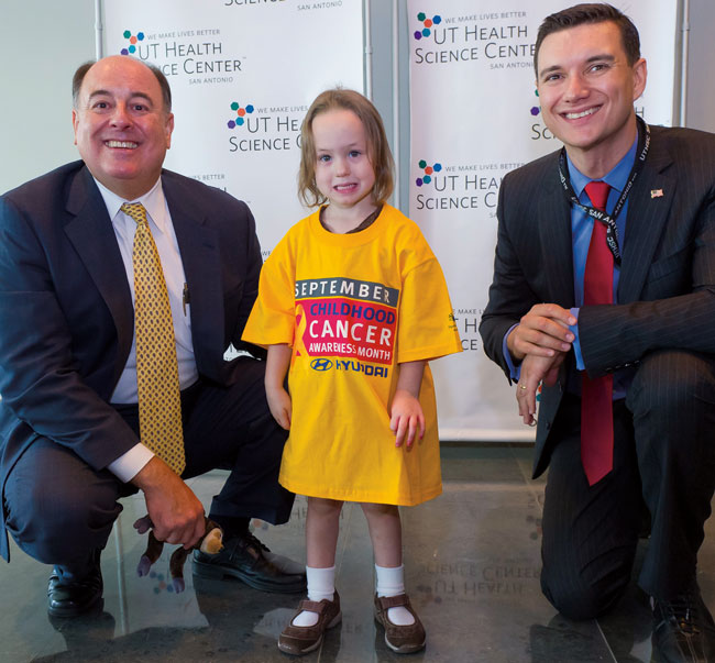
Hyundai Motor America and local Hyundai dealers brought the Hyundai Hope on Wheels™ program to San Antonio, presenting a $250,000 Hope Grant to scientists in the School of Medicine at the UT Health Science Center San Antonio.
The Hope Grant will support studies of myelodysplasia and leukemia conducted by Alexander Bishop, D.Phil., and Vivienne Rebel, M.D., Ph.D., assistant professors in the Department of Cellular and Structural Biology. These scientists work in laboratories at the Greehey Children’s Cancer Research Institute (CCRI), where Hyundai officials presented an oversized check.
Myelodysplastic syndromes are serious blood cell disorders in which the bone marrow does not function normally. Dr. Rebel said the syndromes are difficult to treat, prompting the search for novel ways to address them.
Dr. Bishop studies DNA repair defects in syndromes such as Bloom’s syndrome, a rare inherited disorder that frequently leads to cancer and often displays myelodysplastic syndrome. DNA, the genetic blueprint in cells, undergoes insults and repair constantly. The insults are from environmental and other factors.
"We’re very excited about this grant," Dr. Bishop said. "We asked whether the cells defective in patients with myelodysplasia have DNA repair defects, and the answer is yes. With this grant we can now ask why."
"Everyone knows someone, perhaps a child, who has been touched by cancer," said Gail Tomlinson, M.D., Ph.D., who holds the Greehey Distinguished Chair in the Genetics of Cancer at the UT Health Science Center. "These university-community relationships are so important."

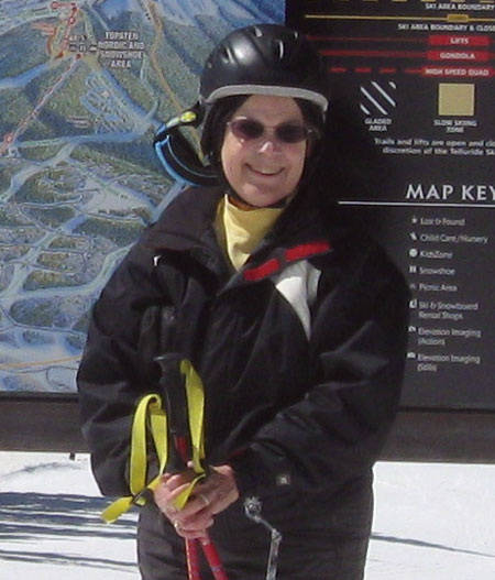
Outsmarting osteoporosis
Virginia Bowden, Ph.D., loves the slopes of Telluride, Colo., where she and her husband, Charles Bowden, M.D., own a condominium. "We’ve been skiing since the 1970s, currently about 30 days a year," she said. "For me the appeal is the mountain views." In 1997 she fractured her hip while skiing at Winter Park. The bone did not heal after the Colorado surgeon inserted a pin, so the following year she underwent a hip replacement at University Hospital in San Antonio. The surgeon sent a bone specimen for clinical testing, which revealed osteoporosis.
Nearly 40 million Americans suffer from osteoporosis or are at high risk for it, according to the National Institutes of Health. This bone-thinning disease is expected to cause fractures that, by 2025, will have cost patients $25 billion to repair. Osteoporosis is most common in post-menopausal Caucasian women, some of whom live their days with the knowledge that a fracture can result from stumping a toe, getting into bed or sneezing. There is great need for translational research to speed scientific discoveries to patients. Two exciting findings in the Long School of Medicine at the UT Health Science Center San Antonio hold new promise to reduce the suffering caused by bone loss.
 Dr. Bowden, director emeritus of the Health Science Center libraries, is a patient of Jan Bruder, M.D., endocrinologist with UT Health Physicians, the clinical practice of the Long School of Medicine. Dr. Bruder sees patients during a weekly osteoporosis consult clinic at the Medical Arts & Research Center. "I’ve been going to Dr. Bruder annually since that time," Dr. Bowden said. "I know what my treatment is, and I think in some ways it was good to find out I had this tendency, so I know to take my calcium, exercise and do the things I should do anyway."
Bone is constantly remodeling — older bone is removed and new bone forms. Exercise places mechanical loads on bone, resulting in a healthy balance of new and old. "The need for regular exercise extends far beyond its role in reducing body fat to maintain a healthy body," said Jean Jiang, Ph.D., professor of biochemistry in the Long School of Medicine and member of the Sam and Ann Barshop Institute for Longevity and Aging Studies. "It is equally important for maintaining healthy bones to avoid osteoporosis and bone loss during aging."
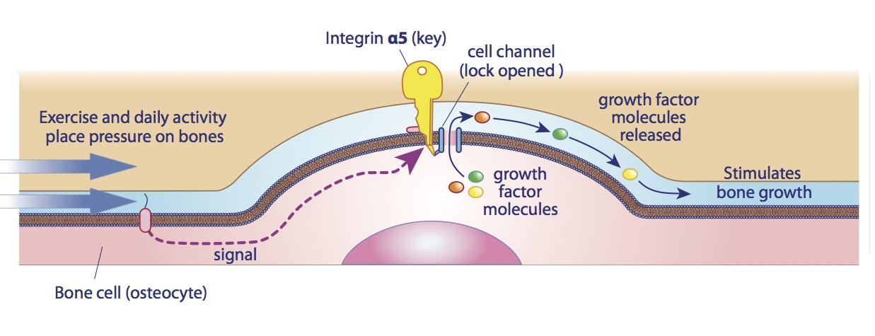
Dr. Jiang’s laboratory discovered novel information about how bones, one, sense the loads being applied to them and, two, release growth factors to build new bone. The report in Proceedings of the National Academy of Sciencesspurred sufficient interest to become an Editor’s Choice article in the journalScience.
Dr. Jiang’s team discovered a lock-and-key system in load-sensing cells. A protein (integrin α5) acts as a key to open the locks, which are channels on the cells’ surface. This releases molecules that trigger bone remodeling. "Our study links the effect of mechanical forces directly to the anabolic (or growth) function of the bone," Dr. Jiang said. "This novel finding could be very useful for development of therapeutic targets to promote the opening of the channels and permit the release of bone growth factors."
Such knowledge could prevent the bone weakening and loss caused by osteoporosis, she said.
Barshop Institute member Brian Herman, Ph.D., is professor of cellular and structural biology in the Long School of Medicine, special assistant to the president of the Health Science Center and a Chancellor’s Health Fellow in Collaboration with The University of Texas System. His team published studies on age-related osteoporosis in mice. The Herman laboratory found that increasing the genetic expression of a protein called caspase 2 prevented osteoporosis development in the rodents. Their bones were 30 percent stronger than the bones of mice lacking caspase 2 expression.
"We are looking at whether caspase 2 can enhance fracture healing," Dr. Herman said. "Our plan is to license technology based on this finding or think about starting a company ourselves."
More-effective, better-tolerated treatments for bone health are needed. Bisphosphonates are the most frequently prescribed medications for osteoporosis. With continuing use, these drugs can cause nausea, abdominal pain, difficulty swallowing and risk of esophageal inflammation and ulcers. In 2010 the National Osteoporosis Foundation issued new treatment recommendations; one was for patients to take drug holidays. "There are side effects of long-term treatment of osteoporosis, and based on bone density and risk factors some people who were previously started on therapy would not be started today," Dr. Bruder, also a Barshop Institute member, said. "This is not to say that patients can go untreated, however. If you need to be on therapy, you need to be on it."
Dr. Bowden served the Health Science Center from 1970 to 2003, including as library director from 1985 to 2003. Her osteoporosis medication is re-evaluated once a year. For three years she received an annual infusion; Dr. Bruder said it was not necessary this year.
The loads from exercise on the Colorado snow continue to strengthen her bones. "I’m still skiing," Dr. Bowden said. "I stick with the intermediate-level areas and try not to fall. I was knocked down this past year on Christmas Eve by a snowboarder and broke my collarbone. The ski patrol came quickly and took me to the clinic. The bone healed within a month and I was skiing again. It (osteoporosis) hasn’t slowed me down."

Clues from clams and hydra
Steven Austad, Ph.D., professor of cellular and structural biology, points to clams in an aquarium at the Sam and Ann Barshop Institute for Longevity and Aging Studies. The institute is part of at the UT Health Science Center at San Antonio. "The lighter ones, typically called ‘hard clams,’ are served in restaurants and live 100 years," he says. "The darker ones, called 'ocean quahogs,' live 400 to 500 years. These clams are older than any other living animal."

It’s a little-known fact that clams have beating hearts. "Its heart beats slower than the human heart but it lives much, much longer," Dr. Austad says. "In fact, it beats as many times as the human heart over its lifetime." To determine the age of a clam, its shell is cut open with a diamond saw and its internal rings are counted. From examining the width of these rings, which is much like reading a bar code, marine biologists study ancient ocean conditions.
In another room, Dr. Austad encourages visitors to peer through a microscope at hydra — small polyps typically found on the underside of aquatic vegetation in freshwater pools and streams. These fascinating invertebrates don’t age and have virtually unlimited regenerative potential. He compares hydra to a villain from one of the "Terminator" movies who, whenever shot or otherwise maimed, pools himself back together. Hydra species can be disassembled into "a pile of cells that remarkably can reassemble themselves into a whole hydra again," Dr. Austad says.
Dr. Austad, interim director of the Barshop Institute, studies the comparative biology of aging.The field asks questions about species because of their exceptionally slow or rapid aging rates. The goal is to understand the unknown and unexplored mechanisms of aging. What causes us to age? What factors, both genetic and environmental, impact the process?
[pexyoutube pex_attr_src="https://www.youtube.com/watch?feature=player_detailpage&v=m5t0KNqM0Zs"][/pexyoutube]
In clams, he is assessing the rate of protein turnover. In hydra, he is inducing the polyps to reproduce, which ends the polyps’ immortal status and sets them on the path of aging. "This tells us which genes are important," Dr. Austad says. In one of his many scientific articles, he writes of the hydra: "Thus, we have the intriguing phenomenon that aging and its absence can potentially both be observed in the same species."
Mexican free-tail bats, selected small nonhuman primates, and birds such as budgerigars, canaries and zebra finches are also under study or are candidates for study. Because of new cellular and molecular techniques for investigating novel species, "the new comparative biology of aging is poised to dwarf earlier contributions," Dr. Austad says.

Preserving memory
 After the 40th birthday it becomes necessary to write notes — and then try to remember the location of the notepad. Learning and memory decline with age, and for some the cliff is Alzheimer’s disease. Thankfully, scientists from the Sam and Ann Barshop Institute for Longevity and Aging Studies at the UT Health Science Center at San Antonio are studying a compound that could rescue our flagging memories.
The researchers added a bacterial product — rapamycin — to the diet of healthy mice throughout their life span. Rapamycin, first isolated from soil on Easter Island, enhanced learning and memory in young mice and improved these faculties in old mice, studies showed.
"We made the young ones learn, and remember what they learned, better than what is normal," said the Barshop Institute’s Veronica Galvan, Ph.D., assistant professor of physiology in the Long School of Medicine. "Among the older mice, the ones fed with a diet including rapamycin had an improvement, negating the normal decline you see in these functions with age."
The drug also lowered anxiety and depression-like behavior in the mice; anxiety and depression are factors that impair human cognitive performance. Jonathan Halloran of the Galvan laboratory conducted scientifically reliable tests to measure these cognitive components in the rodents.
Mice are burrowers and are happy in a tunnel with walls. Halloran used an elevated maze of tunnels leading to a catwalk without walls. "All of a sudden the mice are in open space," Halloran said. "It’s pretty far from the floor for their size, sort of like if you’re hiking and all of a sudden the trail gets steep. It’s pretty far down and not so comfortable."
Mice with less anxiety are more curious to explore the catwalk. "We observed that the mice fed with a diet containing rapamycin spent significantly more time out in the open arms of the catwalk than the animals fed with a regular diet," Halloran said.
The second test measured depression-like behavior in the rodents. Mice do not like to be held by their tails, which is the way they are moved from cage to cage. Inevitably they struggle to find a way out. "How much and how often they struggle is a measure of the motivation they have to get out of an uncomfortable situation," Dr. Galvan said.
Some mice barely struggle to get free, but if an antidepressant is administered they struggle a lot more. "We found rapamycin acts like an antidepressant — it increases the time the mice are trying to get out of the situation," Dr. Galvan said. Anxiety and depression-like behavior decreased in all ages of mice fed with the rapamycin-enhanced diet.
The researchers measured levels of three "happy, feel-good" neurotransmitters: serotonin, dopamine and norepinephrine. All were significantly augmented in the midbrains of mice treated with rapamycin. "This is super-interesting, something we are going to pursue in the lab," Dr. Galvan said.
Rapamycin rescued learning and memory in mice affected by Alzheimer’s-like deficits, the team previously reported. In the new studies, the enhancements are demonstrated even in healthy mice. The elevation of the three neurotransmitters, which are chemical messengers in the brain, may explain how rapamycin accomplished this, Dr. Galvan said.
Rapamycin is an antifungal agent administered to transplant patients to prevent organ rejection. The drug is named for Rapa Nui, the Polynesian title for Easter Island. This island, 2,000 miles from any population centers, is the famed site of nearly 900 monolithic statues (pictured above). If rapamycin proves to be a suitable therapy for human cognition, this mysterious atoll could be called the Easter Island of learning and memory.
[pexyoutube pex_attr_src="https://www.youtube.com/watch?v=ja-3UAAoAUc"][/pexyoutube]

The CTRC A-Team
In the ongoing battle against a deadly disease, the Cancer Therapy & Research Center has assembled the ultimate team of professionals.
 In November 2007 Texas voters passed Proposition 15, the constitutional amendment that established the Cancer Prevention and Research Institute of Texas (CPRIT). The amendment authorizes the issuance of $3 billion in bonds to support the institute, which exists to enhance cancer research, prevention and control programs across Texas.
Thanks to this historic vote, biochemist Dmitri Ivanov, Ph.D., is the inaugural CPRIT Scholar in Cancer Research at the UT Health Science Center San Antonio.
Ask Dr. Ivanov and he will tell you: The excitement of scientific discovery is in his veins.
He grew up in St. Petersburg, Russia, the son of two material scientists. His father, Dr. Nikolay Ivanov, is a plastics expert and his mother, Dr. Raisa Grishmanovskaya, is an expert in stainless steel. "In general, the reason I came into science is that I came from a family of scientists," the younger Ivanov said.
His interest in cancer is personal, given that his father was diagnosed with bladder cancer in 2005 when Dr. Dmitri Ivanov was a postdoctoral research fellow at Harvard Medical School. "That stimulated an interest in using my expertise to fight cancer," he said.
His research examines how cells repair damage to their genetic instructions, called DNA. Cells respond to genetic insults wreaked by environmental factors, ultraviolet radiation in sunlight, and oxidative stress generated by the cells’ own energy consumption. Efficient DNA repair should be a good thing, but tumor cells take advantage of it for nefarious purposes, namely to grow. "What intrigues us is there are a lot of very intimate connections between DNA repair pathways and signaling pathways that are deregulated in cancer," Dr. Ivanov said.
Thanks to the CPRIT Scholar grant, which is $2 million over four years, Dr. Ivanov’s research program in San Antonio is getting a strong start. At Harvard he helped identify a potential target for inhibition of one of the key DNA repair pathways, called nucleotide excision repair. At the Health Science Center he wants to expand into different DNA repair pathways. "In particular we will study the Fanconi anemia pathway," he said. "Fanconi anemia is a rare genetic disorder. People with Fanconi anemia have this DNA repair pathway impaired, and one of the hallmarks of the disorder is high incidence of cancer. We will investigate whether this pathway could be targeted for anti-cancer therapy."
Because cancers can use DNA repair to survive, lowering the amount of repair activity can curb tumor growth while still enabling healthy cells to enact repairs.
Dr. Ivanov used a battlefield analogy: "In combat you don’t want to explode a big bomb that kills your enemy but kills your own troops, too. You want your bomb to go to a specific location or not go to a specific location. Similarly, we would like to be more discriminate in the way we block protein function. We don’t want the bomb to go where our own troops, aka the healthy cells, are."
Dr. Ivanov has an ambitious research agenda and is willing to take risks on new ideas. "The CPRIT program really gives us the luxury to be innovative and try new things," he said. "When you are a young scientist, you are more likely to take the unexpected route. CPRIT’s support is an invaluable resource for scientists at the early stages of their careers."
Bruce J. Nicholson, Ph.D., professor and chairman of biochemistry in the Graduate School of Biomedical Sciences, spoke about the team of scientists in San Antonio working on discoveries that have potential to be carried into clinical trials.
"Dr. Ivanov’s arrival is a key component of our plan to build a team of biochemists who will provide essential insights into the structure of multi-molecular complexes essential to the control of many cell processes. The knowledge of these structures is being used in the development of anti-cancer agents that can be tested in Phase I clinical trials [first-time use in humans] at the Cancer Therapy& Research Center at the UT Health Science Center.
"Coordinated with expanding efforts in medicinal chemistry and high-throughput screening in collaboration with The University of Texas at San Antonio and Southwest Research Institute, drug discovery is undergoing a rebirth in San Antonio that promises exciting prospects for the near future."
Breastbone mechanics
UT Medicine surgeons revolutionize repair of unstable breastbones
Using a "mixed tool bag of techniques," including a ratchet mechanism, UT Health San Antonio physicians in three disciplines — cardiothoracic surgery, radiology and plastic surgery — provide a new breakthrough for patients with unstable breastbones.
Leroy Lorenz’s breastbone failed to heal back together after a coronary artery bypass operation. Donna Lea Anderson’s breastbone was eaten away by a staph infection, a complication of aortic valve surgery. Both lived with chronic pain and disconcerting internal movement for months. Then they discovered an innovative breastbone reconstruction technique performed by specialists from UT Health Physicians. UT Medicine is the clinical practice of the Long School of Medicine at the UT Health Science Center.
Leroy Lorenz and Donna Lea Anderson are among the estimated 400,000 people a year who undergo surgery to bypass arteries, repair heart valves and correct other problems. These surgeries require dividing the breastbone, which afterward is commonly repaired with stainless steel wire. Healing is normal in most cases, but in up to 3 percent of patients (12,000 a year) the breastbone comes apart. Morbid obesity, poorly controlled diabetes, chronic coughing and vitamin D deficiency often related to osteoporosis increase the risk of this serious complication.
The breastbone, or sternum, anchors the rib cage protecting the heart and lungs. Cardiothoracic surgeons, plastic surgeons and radiologists from UT Medicine pioneered a corrective surgery that employs three technologies to help these patients.

A year after his surgery, Leroy Lorenz’s reconstructed breastbone is strong enough to handle the jostling of mowing the family property on his new lawn tractor. He’s also trimming oak trees with a pole trimmer, picking up limbs and clearing fence line. It’s a newfound freedom, his wife says.
Cardiothoracic surgeons ensure the heart is not nicked, as its proximity to the breastbone is razor thin. , professor and chair of the Department of Cardiothoracic Surgery in the Long School of Medicine, leads members of a team that work on these high-risk cases. "It is usually quite uncomfortable for a person to not have a solid breastbone," Dr. Calhoon said. "The grating movement of an incompletely healed sternum can cause chronic pain and lead to anxiety and depression. These patients are often led to believe nothing can be done."
Cardiothoracic surgeons ensure the heart is not nicked, as its proximity to the breastbone is razor thin. , professor and chair of the Department of Cardiothoracic Surgery in the Long School of Medicine, leads members of a team that work on these high-risk cases. "It is usually quite uncomfortable for a person to not have a solid breastbone," Dr. Calhoon said. "The grating movement of an incompletely healed sternum can cause chronic pain and lead to anxiety and depression. These patients are often led to believe nothing can be done."
Cardiothoracic surgeons ensure the heart is not nicked, as its proximity to the breastbone is razor thin. John Calhoon M.D., professor and chair of the Department of Cardiothoracic Surgery in the Long School of Medicine, leads members of a team that work on these high-risk cases. "It is usually quite uncomfortable for a person to not have a solid breastbone," Dr. Calhoon said. "The grating movement of an incompletely healed sternum can cause chronic pain and lead to anxiety and depression. These patients are often led to believe nothing can be done."
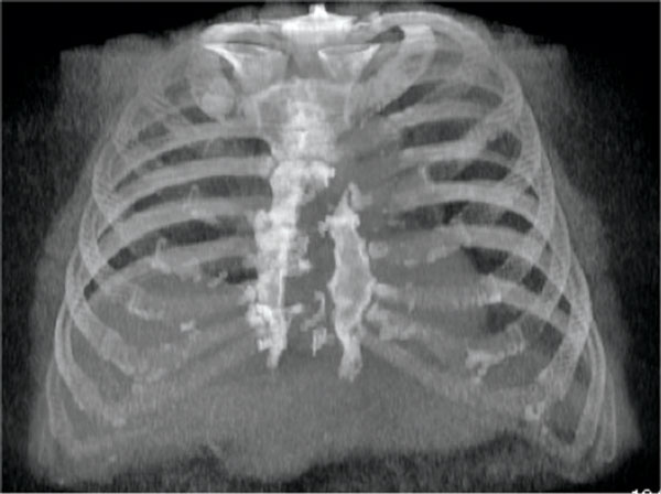
Lorenz, a truck driver from Seguin, underwent reconstruction in May 2011 at CHRISTUS Santa Rosa Hospital - New Braunfels. His breastbone was divided in a bypass operation in 2009. After the surgery, when it was expected to have healed, he felt a gap inside and shared this with his wife, Leigh Ann. "You could actually put your hand on the bone and move it around and feel all my innards moving," he said. A chest X-ray confirmed that the breastbone had separated and the wires were broken into several pieces.
Anderson, a writer/lyricist from San Antonio, underwent reconstruction in April 2011 at University Hospital in San Antonio. A staph infection destroyed her sternum after her second heart valve operation, causing the top four-fifths of the sternum to be removed. "For a year and a half, my ribs rubbed together and I heard popping whenever I moved," she said.
How the surgery is done
Carlos Restrepo, M.D., a chest radiologist with UT Medicine, uses advanced CT imaging to confirm the separation of the sternum. Because every patient is different, it is important to visualize the exact anatomic details surgery will correct. "CT images also show infection, if it is present, which is a common explanation for why the surgical wound is opening instead of healing," Dr. Restrepo said.

 In surgeries such as artery bypass and heart valve repair, the breastbone is divided and afterward is commonly repaired with stainless steel wire (top). In a small percentage of cases, the breastbone comes apart and the wires can break. UT Health San Antonio physicians are using several techniques, including Sternal Talons® illustrated above, to fix the breastbone in place and correct the problem.
Aided by digital imaging, a cardiothoracic surgeon lifts the sternum off the heart and removes scar tissue on it. A UT Medicine plastic surgeon on the case, either Howard Wang, M.D., or Luis Jaramillo, M.D., dissects muscle away from the breastbone. With the breastbone exposed, the cardiothoracic surgical team reassembles the pieces like a puzzle, using a mixed "tool bag of techniques" sometimes including Sternal Talons® to fix the breastbone in place. A ratchet mechanism is used to pull the sides together.
The cardiothoracic surgeon may also utilize orthopedic plating techniques to stabilize the sternum. Because the two sides of the fracture are jagged and leave gaps when drawn tight, the surgeon may also employ Kryptonite™ Bone Cement, which provides a bridge for bone cells to migrate across, filling the gaps. Full healing can then take place.
Afterward, a plastic surgeon inserts muscle or fat from the chest or other parts of the body to fill the chest space with strong tissue that brings good circulation. This provides protection, blood supply, healing and the ability to deliver antibiotics. "Bone, tendon and vital organs must be covered quickly to prevent dangerous infection," Dr. Wang said.
Preventing problems
For high-risk patients such as diabetics who undergo chest surgery, a number of sternal closure methods such as those described above may be considered. "At the end of the day, what matters most is creating a solid breastbone again as safely as possible," Dr. Calhoon said.
"I put up with it (the unnatural feeling in his chest) for a year and a half, but since the operation I’ve been able to do more stuff around home than I have the last two years," Lorenz said.
Anderson’s surgery also involved implantation of synthetic bone to replace the large amount of sternum that deteriorated by infection. "I can move now without hearing the snap, crackle and pop," Anderson said. "I know my sternum is stabilized and mentally that does so much for me."
For information about this type of surgery, call UT Health San Antonio Cardiothoracic Surgery at 210-358-8001 or visit UTHealthPhysicians.org