Lung cancer tumors studied to discover new treatment, increase survival rate
Mays Cancer Center Annual Report
Lung cancer is a leading cause of cancer-related deaths worldwide. Previously, standard treatment for unresectable lung cancer, which cannot be surgically removed, was chemotherapy alone or in combination with radiation.
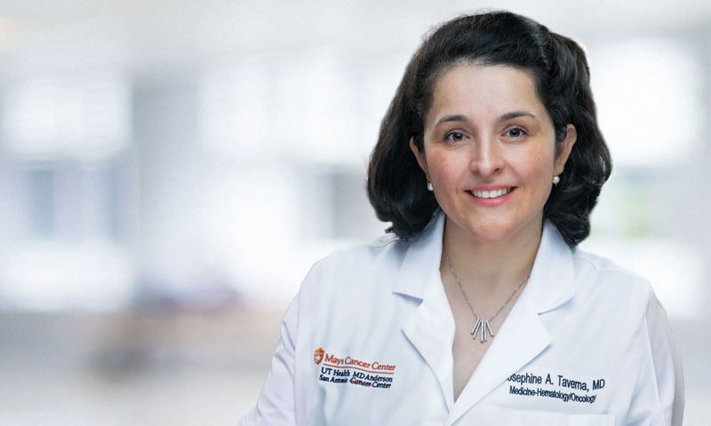
Josephine Taverna, M.D., a physician-scientist at the Mays Cancer Center, said current treatment involves chemotherapy and immunotherapy, a newer treatment using a person’s immune system to fight diseases such as cancer. With the advancement of immunotherapies, the survival outcomes for a patient with metastatic lung cancer (tumors that has spread to distant organs) at five years is 24 percent.
Dr. Taverna, assistant professor of medicine in the Division of Hematology and Oncology at the Long School of Medicine at UT Health San Antonio, is part of a multidisciplinary team performing advanced research with the goal of greatly increasing the survival rate for patients diagnosed with lung cancer.
The Mays Cancer Center is currently conducting early phase clinical trials. Dr. Taverna is collecting patient-derived organoids, also known as “human tissue in a dish,” for drug testing experiments. Lung organoids are lung cancer tumors and surrounding tissue that can be grown in a petri dish to create a living biobank for individual lung cancer patients. These organoids can be used for therapeutic drug discovery and can be used for personalized treatments.
“We have been developing the lung organoid research program since 2018. We have been growing lung tumors outside the human body,” she said. “The goal is to personalize treatment by testing new drugs on these tumors ex vivo (outside the human body). We can combine the individual tumor tissue and the drugs in the petri dish and see the effect of the drug on tumor growth. If the tumor responds, then the drug could be tested in the context of a clinical trial. This would allow us to provide personalized care to each patient.”
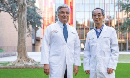
She is working under her mentor, Tim Huang, Ph.D., professor and Alice P. McDermott President’s Distinguished University Chair in Molecular Medicine. Dr. Huang’s lab has been performing genetic analyses of cancer for almost 30 years. His team uses next-generation sequencing technologies for integrative analysis of cancer genomes.
“I’ve been working side by side with Dr. Huang in his lab. We are taking the human lung tumor and studying protein expression at a single-cell level to study signaling pathways that are active not only in the lung tumor single cells but also immune cells surrounding the tumor,” Dr. Taverna explained.
Single-cell profiling is currently being conducted through the Bioanalytics and Single-Cell (BASiC) Core, which was funded by a $3.3 million core facility grant from the Cancer Prevention and Research Institute of Texas (CPRIT) awarded to Dr. Huang. The facility provides standardized, high-quality services to support translational research.
In addition to having cutting-edge equipment, Dr. Taverna emphasizes that the multi-specialty team working on this research has been critical to the success of the lung cancer research.
Daniel DeArmond, M.D., chief of the Division of Thoracic Surgery in the Department of Cardiothoracic Surgery at UT Health, gains approval from his patients, who are newly diagnosed with lung cancer, to provide the tumors and surrounding tissue to the researchers involved in this clinical trial. He collects the tumor and surrounding cells so they can be processed in the lab. The tumor specimen is cultured as organoids in the South Texas Research Facility.
Archival tumor specimen will be examined by pathologists Kenneth Hughes, M.D., and Marjorie David, M.D., Pawel Osmulski, Ph.D., and Maria Gaczynska, Ph.D., are biophysicists studying how the circulating tumor cells metastasize within the blood stream.
Nameer Kirma, Ph.D., director of BASiC Core, oversees the single-cell profiling of tumors using mass cytometry by-time-of-flight (CyTOF) to measure multiple cellular proteins at the single-cell level. CyTOF has allowed the team to identify the AXL and JAK cancer pathways as promising therapeutic targets for lung cancer.
“These oncogenic signaling pathways are critical because they allow us to develop combination treatments for our patients,” she added. “We are now focusing our efforts on lung tumors where both AXL and JAK signals are upregulated,” she said. “We’ve learned that the AXL and JAK pathway is active in most advanced tumors. While we can’t look at a patient and decide how to target therapy, we can look at the individual tumors to see if AXL-JAK signaling is present.”
The next step is designing a clinical trial for patients whose tumors show AXL and JAK upregulated, Dr. Taverna said. “If you can pre-select patients who have a certain biomarker (like AXL or JAK) and treat with a targeted therapy, we are seeing that patients may have more robust treatment responses and improved survival outcomes. In this way, we will be able to develop biomarker-driven clinical trials and accelerate FDA approval for these novel drugs.” Based on this research’s preliminary studies, the next step is to design a Phase II clinical trial with these therapies designed to inhibit the spread of cancer.
“We are at a very exciting time in this field with the advent of organoids and single-cell profiling technologies. These advances will help us select the right therapies or combination therapies for our individual patients. We need to work as quickly as we can so we can help patients diagnosed with lung cancer.”