Mapping the Brain: The Last Frontier

By Michael Seringer
The 17th century French philosopher René Descartes revolutionized mathematics with the development of the Cartesian coordinates, which mark the position of a point in three-dimensional space.
By the early 1980s, Peter Fox, MD, director of the Research Imaging Institute at UT Health San Antonio, was using Descartes’ coordinate system to create standardized brain mapping methods. Dr. Fox’s quantitative method of brain mapping connected form to function, starting what would become an immense data set that has helped fuel a scientific boom of discovery in the brain.
“I developed this approach, published it and then started lobbying people to adopt it,” he said. “And they did!”
Dr. Fox knew early on in his training as a neurologist that a standardized, detailed guide to brain mapping was needed.
“When I was in medical school, I knew I wanted to do functional brain mapping, and there were no tools to do that in radiology,” he said. “The technology hadn’t been invented yet.”
In 1991, Dr. Fox came to UT Health San Antonio to serve as the director of the Research Imaging Institute. His focus on employing leading technology and the pursuit of brain science defines the work being done at UT Health San Antonio and is expanding our understanding of how neurological diseases affect the human brain.
Upending 100 Years of Orthodoxy
For over 100 years, medical orthodoxy assumed that when a person moved, thought, listened, tasted, talked or performed any other tasks, their metabolic rate would go up and blood flow would increase.
“Early on we looked into whether this was true or not. We expected it to be true,” Dr. Fox said. “We were making tools that could very accurately measure blood flow, blood volume, oxygen metabolism, glucose metabolism—these tools were coming online right then.”
Dr. Fox took advantage of positron emission tomography (PET) scan technology to reveal the metabolic function of the brain. As he developed methods to map brain functionality, his investigation showed that while the brain is energy-dependent, any extra tasks do not change its energy demand. The brain is remarkably efficient.
“What we found was really startling,” he said. “We found that oxygen metabolism and glucose metabolism didn’t do the same thing. Blood flow and glucose both went up but not oxygen metabolism. There was disassociation that occurred. The extra brain work you do when you’re doing an active task is not very energy dependent.”
Dr. Fox’s unexpected findings motivated other labs to test his conclusion, resulting in even greater imaging innovation. Teams began investigating using functional MRI (fMRI) to map the brain. The results from the fMRI studies confirmed what the PET scan studies completed by Dr. Fox years earlier had shown—but more importantly, they ushered in the more efficient fMRI technology to map the brain.
Attracting Top Researchers
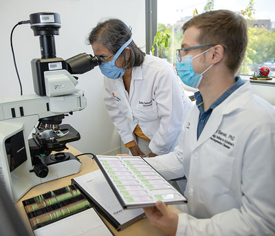
Sudha Seshadri, MD, founding director of the Glenn Biggs Institute for Alzheimer’s & Neurodegenerative Diseases at UT Health San Antonio, partially credits UT Health’s renowned Research Imaging Institute directed by Dr. Fox as one of the reasons she came to San Antonio from Boston.
“To find the earliest stages of the disease—whether the disease is Alzheimer’s or a vascular disease—brain imaging gives us the clues whether there is amyloid in the brain or tau in the brain, whether the brain is showing very subtle signs of a vascular injury, and clearly we want to pick things up in an early stage and keep it from progressing,” she said.
Dr. Seshadri credits the collaborative approach at UT Health San Antonio, in particular with the Research Imaging Institute, as a reason why so many clinical trials are possible at UT Health. The support provided by the imaging research institute is critical to the success of trials investigating Alzheimer’s and dementia.
“We are able to leverage the fact that there is a research imaging institute to attract top talent,” Dr. Seshadri said. “This is expanding not just our data but worldwide data to find the subtlest, earliest signs of different types of brain atrophy and vascular brain injury.”
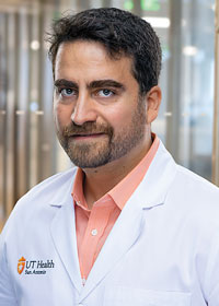
Mohamad Habes, PhD, director of the Biomedical Image Analytics Division at the Research Imaging Institute, said he was very excited to work with Dr. Fox, a renowned leader in the field of brain imaging and mapping. Dr. Fox’s track record of innovation attracted Dr. Habes to come to San Antonio to build advanced analytics and machine learning methods to better predict diseases such as Alzheimer’s and capture related subtle brain changes and injury.
Big Data and the Brain
Dr. Habes is on a mission to obtain as much brain scan data as possible. The more data he funnels into his HIPAA-compliant supercomputer, the more accurate its predictions. Massively large data sets continually improve the machine-learning algorithms built by Dr. Habes. Better artificial intelligence algorithms can predict brain disease earlier and provide more precise treatments.
“The machine-learning algorithms consuming different types of scans will be able to accurately determine how long until a person gets the disease and will be able to provide an individualized guide to treatment,” Dr. Habes said.
By coming to work with Dr. Fox, Dr. Habes gained access to a sufficiently large data set to test his analytics and machine-learning methods. Since the 1980s, Dr. Fox has been collecting and organizing a brain scan database with records from more than 300,000 people. As a past investigator for the Framingham study, the oldest and largest population-based cohort in the United States, Dr. Seshadri is very familiar with large data sets leading to discovery of new medical principles. She points to UT Health San Antonio’s use of sophisticated data science and supercomputing to study neural networks as an example of how large datasets make population studies possible. Investigating neural networks demonstrates a link between the brain’s functional organization and disease—further proof of the power of data analytics in driving brain research.
“When you get schizophrenia, depression or really any disease, it follows the same network patterns that are present in the healthy brain,” Dr. Fox said. “The network patterns that we see in health are being affected selectively by disease. You need a lot of data to see these subtleties.”
Advances in brain imaging and data science pioneered by Dr. Fox are largely responsible for the huge growth in brain mapping research. Over the last 20 years, Dr. Fox has been one of the top-cited neuroscientists in the world. His founding of and participation in various brain mapping organizations demonstrates his commitment to collaboration and fundamental science.
The Research Imaging Institute has grown its faculty and pipeline of research significantly in the last five years. This growth, combined with new innovations in imaging and artificial intelligence, will benefit the health of communities in South Texas. The institute is dedicated to including data from underserved populations that are disproportionately impacted by disease. Unfortunately, minority populations in the United States have lacked meaningful representation in past research, Dr. Seshadri said. UT Health San Antonio is positioned to fill that gap and overcome the existing racial bias on the way to fulfilling the promise of its research.
“I think the next decade will be the time to deliver on the promise of the science. There are so many new ways at looking at these brain diseases that I am hopeful over the next 10 years we are going to see significant breakthroughs,” Dr. Seshadri said.
And when breakthroughs do come for diseases such as Alzheimer’s, dementia, schizophrenia, Parkinson’s and stroke, the research behind their cures will have been made possible by the innovative exploration of the brain by Dr. Fox.