In Alzheimer’s disease, brain cells die. New research shows that their death is linked to disruption of a skeleton that surrounds the nucleus of the cells, a finding that is expected to open new studies on how to prevent the earliest biological events that result in the disease.
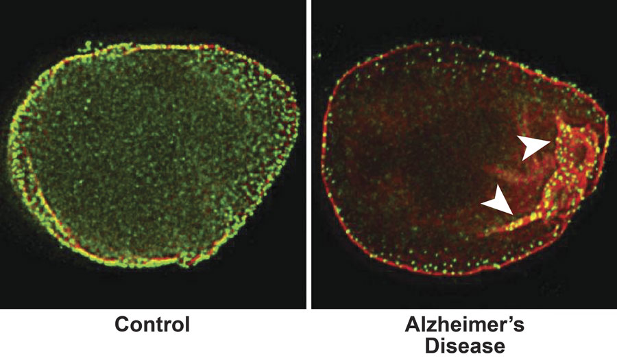
The nucleus is the control center of cells. A mesh-like scaffold called the lamin nucleoskeleton acts like a shield, surrounding and protecting it. But in Alzheimer’s, this skeleton is disordered, said Bess Frost, Ph.D., assistant professor of cellular and structural biology in the Long School of Medicine.
This dysfunction can cause the death of the brain cells. The discovery first was made in a fruit fly disease model. It was confirmed in postmortem brain tissue of people who had Alzheimer’s disease, whose families had donated their brains to research.
“Human brain donation is a very critical part of this work,” Dr. Frost said. “It was important to show that what we found in the fly is really relevant to human disease.”
Dr. Frost and her colleagues at Harvard and Brigham and Women’s Hospital used a technique called super-resolution microscopy to analyze the fruit fly and human specimens. They found peculiar features that looked like tunnels in the lamin of Alzheimer’s-affected specimens.
The team also studied a fruit fly model of Huntington’s disease and did not find any problems with the lamin.
“So, at least compared to one other neurodegenerative disease, lamin dysfunction seems to be specific to Alzheimer’s disease,” she said.
The findings were published in the journal Current Biology.