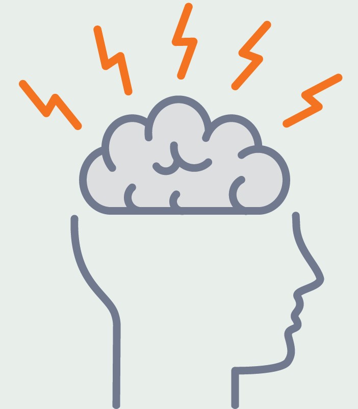
New therapy reduces headache disability after brain injury
The first therapy developed for post-traumatic headache significantly reduced related disability in veterans following a traumatic brain injury and decreased co-occurring symptoms of post-traumatic stress disorder.
The treatment, called Cognitive Behavioral Therapy for Headache, was appealing to patients, showing low drop-out rates. In addition, it is easy for therapists to learn and deliver. The findings were reported in JAMA Neurology.
“We are excited by this development in the treatment of post-traumatic headache, which along with TBI is poorly understood and for which treatment options are so limited,” said Don McGeary, PhD, associate professor of psychiatry and behavioral sciences in the Joe R. and Teresa Lozano Long School of Medicine. “To find the first major treatment success for post-traumatic headache, which is arguably the most debilitating symptom of TBI, and that the treatment also significantly reduces other PTSD symptoms, is a major breakthrough.”
Both TBI and PTSD are signature wounds of post-9/11 military conflicts, and the two conditions commonly occur together. Post-traumatic headaches, or headaches that develop or worsen following a head or neck injury, become chronic and debilitating in a large percentage of those who experience a TBI, such as a concussion, inhibiting their ability to engage in the activities of daily life. When PTSD occurs with other symptoms, it can worsen the headaches and make them more difficult to treat.
Although effective treatments exist for PTSD, they do not for post-traumatic headache. Migraine medications alleviate the headache pain but do not relieve related disability. They also often have unwanted side effects, and their overuse can worsen headaches.
McGeary and his colleagues developed Cognitive Behavioral Therapy for Headache by modifying a psychotherapy for migraine headaches. It includes key components such as relaxation, setting goals for activities patients want to resume, and planning for those situations.
Cognitive Behavioral Therapy for Headache requires eight sessions of 30-45 minutes each. This is shorter than Cognitive Processing Theory, a leading psychotherapy for PTSD that typically requires 12 sessions lasting 60-90 minutes each. The new therapy teaches patients how to evaluate and change upsetting and maladaptive thoughts related to their trauma.
Those receiving Cognitive Behavioral Therapy for Headache reported significant reductions in disability and in negative impact on function and quality of daily life. They also showed improvement in PTSD symptoms. All of these treatment gains were maintained six months after treatment completion. However, the therapy did not reduce headache intensity or frequency compared to usual care, which can include injections, physical and occupational therapies, pain medications, acupuncture and massage, and long-term medical care.
McGeary said the therapy’s reductions on negative life impact are likely due to its building patients’ confidence that they could control or manage their headaches, a concept known as “self-efficacy.” That sense of control was key to helping patients “get their lives back,” he said.
“If you can improve a person’s belief that they can control their headache, they function better,” McGeary said. “That’s because, when dealing with a long-term, disabling pain condition, people make decisions about whether they’re going to actively engage in any kind of activity, especially if the activity exacerbates the pain condition. They make those decisions based on their perceptions of their ability to handle their pain.”
McGeary believes the planning component of the therapy is key to improving those perceptions.Another benefit of the therapy is that it requires only two hours to train clinicians to provide the care. That would make it relatively easy to increase the number of therapists available to treat veterans with post-traumatic headache and ease caseloads at clinics.
In the race to solve Alzheimer’s disease, scientists find more needles in the haystack
Twenty-one million. That’s the number of genetic variations in the human genome that researchers are sifting to identify patterns predisposing people to Alzheimer’s disease.
Because of international collaboration being advanced by UT Health San Antonio faculty, more genetic variations for Alzheimer’s disease are known today than ever before. The gene variants recognized for late-onset Alzheimer’s grew from one in 2009 to 40 in 2022. Recently, scientists published an expanded list of 75, some of which are considered prime drug targets.
It’s a huge haystack, and Alzheimer’s-related genetic variations, like needles, are minuscule in comparison. Sudha Seshadri, MD, Habil Zare, PhD, and other faculty at the university’s Glenn Biggs Institute for Alzheimer’s and Neurodegenerative Diseases are investigators on a global project to answer the many Alzheimer’s riddles.
Seshadri is a founding principal investigator of the International Genomics of Alzheimer’s Project (IGAP). Biggs Institute faculty contributed data for the newest research from IGAP, published in Nature Genetics, and helped craft the discussion on implications of the findings.
“We are looking for the genetic basis to better understand all the different types of biology that may be responsible for Alzheimer’s disease,” said Seshadri, founding director of the Biggs Institute and professor of neurology in the Joe R. and Teresa Lozano Long School of Medicine. “As we include data from more and more people, we are able to find variants that are fairly rare, that are only seen in about 1% of the population.”
Older Hispanic adults are estimated to be at 1.5 times greater risk of Alzheimer’s and other dementias than non-Hispanic whites.
The South Texas Alzheimer’s Disease Research Center — the only designated center in Texas — is a collaboration of the Biggs Institute and The University of Texas Rio Grande Valley.

Eating fish could make you smarter
Eating cold-water fish and other sources of omega-3 fatty acids may preserve brain health and enhance cognition in middle age, new evidence indicates.
Having at least some omega-3s in red blood cells was associated with better brain structure and cognitive function among healthy study volunteers in their 40s and 50s, according to research published online Oct. 5 in Neurology, the medical journal of the American Academy of Neurology. Faculty of UT Health San Antonio and other investigators of the Framingham Heart Study conducted the analysis.
“Studies have looked at this association in older populations. The new contribution here is that, even at younger ages, if you have a diet that includes some omega-3 fatty acids, you are already protecting your brain for most of the indicators of brain aging that we see at middle age,” said Claudia Satizabal, PhD, assistant professor of population health sciences with the Glenn Biggs Institute for Alzheimer’s and Neurodegenerative Diseases at UT Health San Antonio. Satizabal is the lead author of the study.
Volunteers’ average age was 46. The team looked at the relation of red blood cell omega-3 fatty acid concentrations with MRI and cognitive markers of brain aging. Researchers also studied the effect of omega-3 red blood cell concentrations in volunteers who carried APOE4, a genetic variation linked to higher risk of Alzheimer’s disease.
The study of 2,183 dementia- and stroke-free participants found that:
- A higher omega-3 index was associated with larger hippocampal volumes. The hippocampus, a structure in the brain, plays a major role in learning and memory.
- Consuming more omega-3s was associated with better abstract reasoning, or the ability to understand complex concepts using logical thinking.
- APOE4 carriers with a higher omega-3 index had less small-vessel disease. The APOE4 gene is associated with cardiovascular disease and vascular dementia.
The team divided participants into those who had very little omega-3 red blood cell concentration and those who had at least a little.
“We saw the worst outcomes in the people who had the lowest consumption of omega-3s,” Satizabal said. “So, that is something interesting. Although the more omega-3 the more benefits for the brain, you just need to eat some to see benefits.”
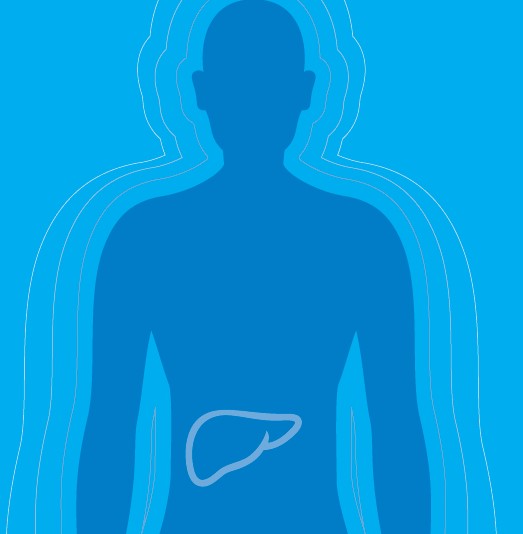
Losing weight could come down to your liver
Losing weight could come down to your liver
 In a breakthrough finding, UT Health San Antonio scientists discovered that inhibiting a liver enzyme in obese mice decreased the rodents’ appetites, increased energy expenditure in fat tissues and resulted in weight loss.
The finding, published in Cell Metabolism, provides a potential drug target to treat metabolic issues such as obesity and diabetes.
“We first needed to discover this mechanism and, now that we have, we can develop drugs to improve metabolic syndrome,” said senior author Masahiro Morita, PhD, assistant professor of molecular medicine in the university’s Sam and Ann Barshop Institute for Longevity and Aging Studies.
“We have an enzyme inhibitor that we want to make more specific to increase its effects,” said first author Sakie Katsumura, DDS, PhD, postdoctoral fellow in the Morita laboratory.
They targeted a liver enzyme called CNOT6L deadenylase. It turns off messenger ribonucleic acids, called mRNAs, that ordinarily carry genetic instructions from the nucleus to sites in the cell where two liver proteins are made. One protein sends signals to the brain to control food intake. The other sends signals to adipose tissues to increase energy expenditure.
The liver enzyme impedes these signals, reducing the benefits. So, the researchers created a first-in-class CNOT6L inhibitor, dubbed iD1, to stabilize the messenger ribonucleic acids in obese mice and increase levels of the two proteins in the blood. After 12 weeks, treated rodents ate less food and showed 30% reduced body weight. Energy expenditures in the adipose tissues increased by 15%. Liver fat decreased 30%.
They also showed improved insulin sensitivity and lower blood glucose levels.
In Texas and the U.S., obesity, Type 2 diabetes, fatty liver disease and related metabolic disorders are at epidemic proportions.
According to the Centers for Disease Control and Prevention, more than 37 million Americans have diabetes. Type 2 diabetes represents at least 90% of the cases. In Texas, approximately 2.7 million people have diagnosed diabetes, and an additional 600,000 people in Texas have diabetes but don’t know it. Another 7 million people in Texas have prediabetes.
Obesity prevalence in the U.S. is more than 40% and is climbing, according to the CDC. Obesity-related diseases include heart attack, stroke, Type 2 diabetes and some cancers.
Next, the researchers will refine the mechanism and identify new drugs that may be more specific and more potent, Katsumura said.
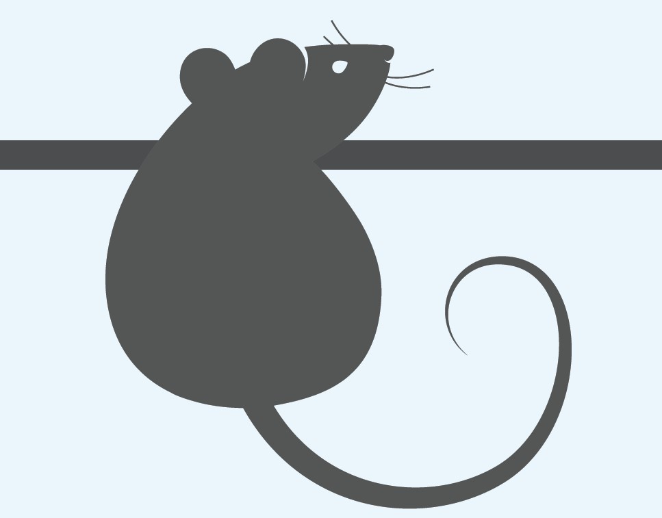
Mouse pups’ cries give clues about autism spectrum disorder
One-fifth of babies who inherit a genetic variant located on chromosome 16 will develop autism spectrum disorder by age 3. The variant is called 16p11.2 deletion.
Noboru Hiroi, PhD, professor in the departments of Pharmacology, Cellular and Integrative Physiology, and Cell Systems and Anatomy, is studying mice that have this deletion. The team is harnessing the power of machine learning to understand which vocalizations of the newborn mouse pups are most predictive of social abnormalities one month later when the pups reach puberty.
“It is essential to identify those very early signs that can predict what is to come, because if we can translate what we discover in mouse pups to human infants and apply therapeutic options earlier, their outcome will be better,” Hiroi said.
When mouse pups are separated from their mothers, they emit ultrasonic vocalizations in a certain order. Mouse mothers respond to the cries and take care of their offspring.
The cries are abnormal in mice with the 16p11.2 deletion.
“Pups that carry this genetic variation cannot form the correct sequence,” Hiroi said. “We want to know whether those abnormal sequences or combinations of call types can predict what is to come one month later in their social behaviors.”
Machine learning will enable the team to develop a precise diagnostic algorithm for autism spectrum disorder.
“Once we can do this with the mouse vocalizations, we can apply the same algorithm to the cries of human babies,” Hiroi said.
Infants at risk of the disorder who are identified in this way can be given desensitization therapy so that they don’t overreact to certain cues they don’t like, he said. And behavioral therapies can be applied to help babies learn how to cope in social situations.
The research is described in the journal Molecular Psychiatry.

‘It’s like being able to see a dime on the surface of the moon.’
When physicians observe or hear of symptoms in their patients, they order CT scans, MRIs or other types of imaging analyses to understand the causes.
Clinical imaging, however, does not illuminate the vast machinations of life that occur deeper within, at the Lilliputian level of individual proteins and other molecules. Chain reactions in this tiny domain determine whether good health continues, or diseases begin. To explore how the reactions foster disorder, scientists acquire images with state-of-the-art instruments capable of ultra-high resolution. They employ statistical models to work out molecular structures. The overall analysis yields sites of disease susceptibility that can be targets for drug therapies.
UT Health San Antonio is investing $5 million over the next three years in such a technology, called cryo-electron microscopy, or cryo-EM for short.
“We are essentially molecular photographers,” said Shaun Olsen, PhD, associate professor of biochemistry and structural biology and director of structural biology cores. “Some people take pictures of buildings. We take pictures of proteins and want to see what they look like in three dimensions.”
Cryo-EM visualizes proteins that are extremely difficult to image using other techniques, said Elizabeth Wasmuth, PhD, assistant professor of biochemistry and structural biology.
Cryo-EM is complementary to existing structural biology technologies at UT Health San Antonio. X-ray crystallography, for example, exposes a protein crystal to X-rays, diffracting the X-ray beam in directions according to the protein’s structure. Nuclear magnetic resonance (NMR) spectroscopy, meanwhile, demonstrates behavior of an atom nucleus when it is placed in a powerful magnetic field. Experts can infer structure from the behavior they observe.
“We look at molecules in levels of detail that are unparalleled,” Wasmuth said. “So, basically, it’s like being able to see a dime on the surface of the moon. This is the level of resolution that the techniques and tools of structural biology allow us to see about molecules inside cells, inside our bodies. Cryo-EM adds a powerful new dimension to our other methods.”
Some protein targets are too small to be visualized by existing techniques or have flexible, wiggly regions that impede the crystal formation, Wasmuth said. Cryo-EM flash-freezes proteins on thin layers of ice within milliseconds and barrages them with electron beams, generating biologically useful information.
“Having a cryo-EM system will allow us to observe drug targets that couldn’t be visualized by the other methods,” Wasmuth said. “The second week the cryo-EM facility became functional, we were able to solve the structure of a complex of proteins involved in DNA damage repair at impressively high resolution. I am confident that this tool is going to transform structural biology research here like we haven’t seen before.”
Much like the space telescopes in Chile, Hawaii and West Texas are indispensable shared resources for astronomers, cryo-EMs are invaluable shared resources for structural biologists.
“We have to remain competitive, and this is a question that a lot of academic medical centers are trying to decide right now: Where will we fit within the research environment?” said Jennifer Sharpe Potter, PhD, vice president for research. “This acquisition reflects our commitment to making San Antonio a biomedical hub for the United States and the world. The types of visualizations and the questions that this technology advances are investments in the long-term improvement of human health.”

Robotic kidney cancer surgery shows desirable outcomes in study
Kidney cancer is not always confined to the kidney. In advanced cases, this cancer invades the body’s biggest vein, the inferior vena cava, which carries blood out of the kidneys back to the heart. Via the IVC, cancer may infiltrate the liver and heart.
The Mays Cancer Center at UT Health San Antonio is one of the high-volume centers in the U.S. with surgical expertise in treating this serious problem.
In a study featured on the cover of the Journal of Urology, researchers from the Mays Cancer Center and the Department of Urology at UT Health San Antonio show that robotic IVC thrombectomy — the removal of cancer from the inferior vena cava — is not inferior to standard open IVC thrombectomy and is a highly safe and effective alternative approach. The affected kidney is removed along with the tumor during surgery, which is performed at UT Health San Antonio’s clinical partner, University Hospital.
Harshit Garg, MD, urologic oncology fellow in the Department of Urology, is first author of the study, and Dharam Kaushik, MD, urologic oncology fellowship program director, is the senior author.
The open surgery requires an incision that begins 2 inches below the ribcage and extends downward on both sides of the ribcage.
“It looks like an inverted V,” Kaushik said. Next, organs that surround the IVC, such as the liver, are mobilized, and the IVC is clamped above and below the cancer. In this way, surgeons gain control of the inferior vena cava for cancer resection.
“Open surgery has an excellent success rate, and most cases are performed in this manner,” Kaushik said. “But now, with the robotic approach, we can achieve similar results with smaller incisions. Therefore, we need to study the implications of utilizing this newer approach.”
The study is a systematic review and meta-analysis of data from 28 studies that enrolled 1,375 patients at different medical centers. Of these patients, 439 had robotic IVC thrombectomy and 936 had open surgery. Kaushik and his team collaborated with Memorial Sloan Kettering Cancer Center, New York; Cedars-Sinai Medical Center, Los Angeles; and the University of Washington, Seattle, to perform this study.
The results are encouraging, finding:
- Fewer blood transfusions: 18% of robotic patients required transfusions compared to 64% of open patients.
- Fewer complications: 14.5% of robotic patients experienced complications such as bleeding compared to 36.7% of open thrombectomy patients.
These large, technically challenging surgeries last eight to 10 hours and involve a multidisciplinary team of vascular surgeons, cardiac surgeons, transplant surgeons and urologic oncology surgeons, Kaushik said.
This is the largest study to analyze the outcomes of robotic versus open IVC thrombectomy, Kaushik said. While open surgery remains the gold standard for surgery, the study shows the robotic variation could be a good option for certain patients, he added.
“Optimal candidacy for a robotic surgery should be based on a surgeon’s robotic expertise, the extent and burden of the tumor, and the patient’s comorbid conditions,” Kaushik said.