Ewing Halsell Foundation enhances Human Anatomy Program with $500,000 gift
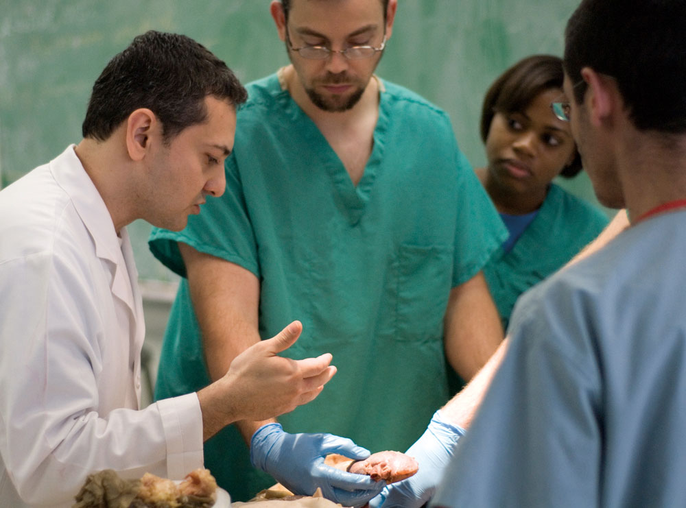
Impacting patients and making lives better are the goals every student at the UT Health Science Center aspires to. That’s why students in all areas of study – medical, dental and health professions – begin their educational journey by meeting their very first patients in human anatomy laboratories on campus.
“Human anatomy introduces students to a different kind of patient,” said Omid Rahimi, Ph.D., assistant professor in the Department of Cellular and Structural Biology, and director of the Human Anatomy Program at the Health Science Center.
“Before students work with living patients, they work with cadavers. These are the remains of once vibrant persons in the midst of life,” Dr. Rahimi said. “These body donors are the preceptors for each student to confront disease, death and dying. They teach students to become skilled and compassionate health care professionals so they can care for their future patients. This is a unique experience that students can’t learn anywhere else; not from a textbook or any one model alone.”
Thanks to a $500,000 gift from the Ewing Halsell Foundation, the students’ human anatomy learning experience at the Health Science Center will be enhanced by new state-of-the-art technology.
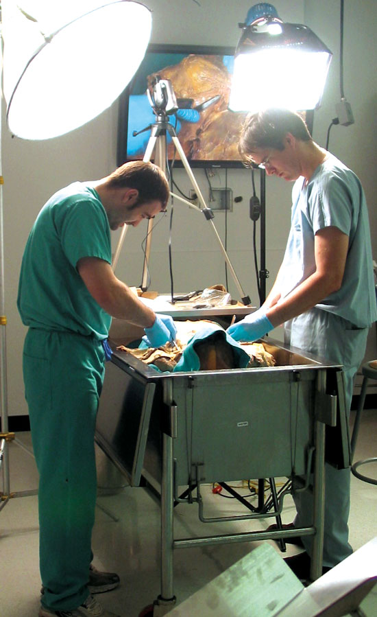
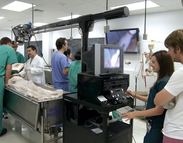
The gift is providing for 25 high-definition 58-inch plasma flat-screen monitors and ceiling-mount speakers. In addition, it also is funding a wireless microphone system, high-definition video cameras, including several hand-held models, a remote-controlled video camera mounted on a telescoping boom arm, as well as tank-side computer systems for the 57 cadaver tanks. All equipment will be networked throughout the four human anatomy laboratories located on the first floor of the Dental School. Installation of the plasma screens began this spring and is expected to be completed and ready for use by faculty and students this fall. Approximately 400 medical, dental, health professions and graduate students benefit from the Human Anatomy Program each year.
Christi A. Walter, Ph.D., professor and chair of the Department of Cellular and Structural Biology, conceived the vision a few years ago to update the labs with her colleagues, including Ron Philo, Ph.D., a retired faculty member and current adjunct associate professor who served as director of the Human Anatomy Program from 2003 to 2010.
“We are so grateful to the trustees of the Ewing Halsell Foundation for their generosity, foresight and dedicated support to the art and science of anatomy education,” Dr. Walter said. “They truly understand the vast impact that anatomy education has on our students’ lives. By enhancing the students’ learning experience, their gift is helping us to better prepare students to care for thousands of patients in the future after they graduate and work in their field.”
Dr. Rahimi added that in addition to improving the students’ learning experience, the new technology will also improve the anatomy faculty members’ teaching capabilities and reach.
“Today, we have a shortage of anatomy faculty,” he said. “Many are retiring and the pipeline of graduate students who solely want to teach anatomy is thin. Faculty members today have to do more with less. Thanks to this gift, we will be able to help fill that void.”
Dr. Rahimi explained how live dissections performed by students at one tank, for example, can be captured by video camera and broadcast to all four laboratories, classrooms or to off-campus sites simultaneously. “All of the bodies are different. So if one group of students discovers an important variation in a particular organ in one cadaver, they can share it in real time with all of their classmates and all of the laboratories,” he said.
The tank-side computers will allow faculty and students to access the Internet or draw from existing human anatomy software, such as the digitized resources of head and neck vasculature and ocular movements developed by the Health Science Center’s own Frank J. Weaker, Ph.D., associate professor of cellular and structural biology.
“This new technology will allow every participant to be more engaged in academic medicine and practice and to work better together and communicate as teams,” Dr. Rahimi said. “In addition to preparing the next generation of surgeons and other health care providers, I think it could reinvigorate a desire in some of our students to later teach anatomy as well.”
Dr. Rahimi noted that on top of supporting students and faculty at the Health Science Center, the Ewing Halsell Foundation gift also pays tribute to the numerous individuals and families who donate their bodies or the bodies of a loved one to the university’s Human Anatomy Program.
“At the end of the day, it’s all about patients – past, present and future,” Dr. Rahimi said. “The Ewing Halsell Foundation trustees have demonstrated their dedication to students and their deep respect for the silent patients who donate their bodies for the sake of science education and patients in the future. We are sincerely thankful.”